Alberto Díez-Montiel, Alicia Pose-Díez-de-la-Lastra, Alba González-Álvarez, José I Salmerón, Javier Pascau, Santiago Ochandiano
{"title":"Tablet-based Augmented reality and 3D printed templates in fully guided Microtia Reconstruction: a clinical workflow.","authors":"Alberto Díez-Montiel, Alicia Pose-Díez-de-la-Lastra, Alba González-Álvarez, José I Salmerón, Javier Pascau, Santiago Ochandiano","doi":"10.1186/s41205-024-00213-2","DOIUrl":null,"url":null,"abstract":"<p><strong>Background: </strong>Microtia is a congenital malformation of the auricle that affects approximately 4 of every 10,000 live newborns. Radiographic film paper is traditionally employed to bidimensionally trace the structures of the contralateral healthy ear in a quasi-artistic manner. Anatomical points provide linear and angular measurements. However, this technique proves time-consuming, subjectivity-rich, and greatly dependent on surgeon expertise. Hence, it's susceptible to shape errors and misplacement.</p><p><strong>Methods: </strong>We present an innovative clinical workflow that combines 3D printing and augmented reality (AR) to increase objectivity and reproducibility of these procedures. Specifically, we introduce patient-specific 3D cutting templates and remodeling molds to carve and construct the cartilaginous framework that will conform the new ear. Moreover, we developed an in-house AR application compatible with any commercial Android tablet. It precisely guides the positioning of the new ear during surgery, ensuring symmetrical alignment with the healthy one and avoiding time-consuming intraoperative linear or angular measurements. Our solution was evaluated in one case, first with controlled experiments in a simulation scenario and finally during surgery.</p><p><strong>Results: </strong>Overall, the ears placed in the simulation scenario had a mean absolute deviation of 2.2 ± 1.7 mm with respect to the reference plan. During the surgical intervention, the reconstructed ear was 3.1 mm longer and 1.3 mm wider with respect to the ideal plan and had a positioning error of 2.7 ± 2.4 mm relative to the contralateral side. Note that in this case, additional morphometric variations were induced from inflammation and other issues intended to be addressed in a subsequent stage of surgery, which are independent of our proposed solution.</p><p><strong>Conclusions: </strong>In this work we propose an innovative workflow that combines 3D printing and AR to improve ear reconstruction and positioning in microtia correction procedures. Our implementation in the surgical workflow showed good accuracy, empowering surgeons to attain consistent and objective outcomes.</p>","PeriodicalId":72036,"journal":{"name":"3D printing in medicine","volume":"10 1","pages":"17"},"PeriodicalIF":3.1000,"publicationDate":"2024-05-31","publicationTypes":"Journal Article","fieldsOfStudy":null,"isOpenAccess":false,"openAccessPdf":"https://www.ncbi.nlm.nih.gov/pmc/articles/PMC11140883/pdf/","citationCount":"0","resultStr":null,"platform":"Semanticscholar","paperid":null,"PeriodicalName":"3D printing in medicine","FirstCategoryId":"1085","ListUrlMain":"https://doi.org/10.1186/s41205-024-00213-2","RegionNum":0,"RegionCategory":null,"ArticlePicture":[],"TitleCN":null,"AbstractTextCN":null,"PMCID":null,"EPubDate":"","PubModel":"","JCR":"Q1","JCRName":"RADIOLOGY, NUCLEAR MEDICINE & MEDICAL IMAGING","Score":null,"Total":0}
引用次数: 0
Abstract
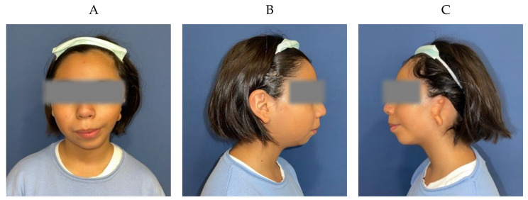
Background: Microtia is a congenital malformation of the auricle that affects approximately 4 of every 10,000 live newborns. Radiographic film paper is traditionally employed to bidimensionally trace the structures of the contralateral healthy ear in a quasi-artistic manner. Anatomical points provide linear and angular measurements. However, this technique proves time-consuming, subjectivity-rich, and greatly dependent on surgeon expertise. Hence, it's susceptible to shape errors and misplacement.
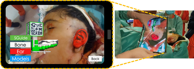
Methods: We present an innovative clinical workflow that combines 3D printing and augmented reality (AR) to increase objectivity and reproducibility of these procedures. Specifically, we introduce patient-specific 3D cutting templates and remodeling molds to carve and construct the cartilaginous framework that will conform the new ear. Moreover, we developed an in-house AR application compatible with any commercial Android tablet. It precisely guides the positioning of the new ear during surgery, ensuring symmetrical alignment with the healthy one and avoiding time-consuming intraoperative linear or angular measurements. Our solution was evaluated in one case, first with controlled experiments in a simulation scenario and finally during surgery.
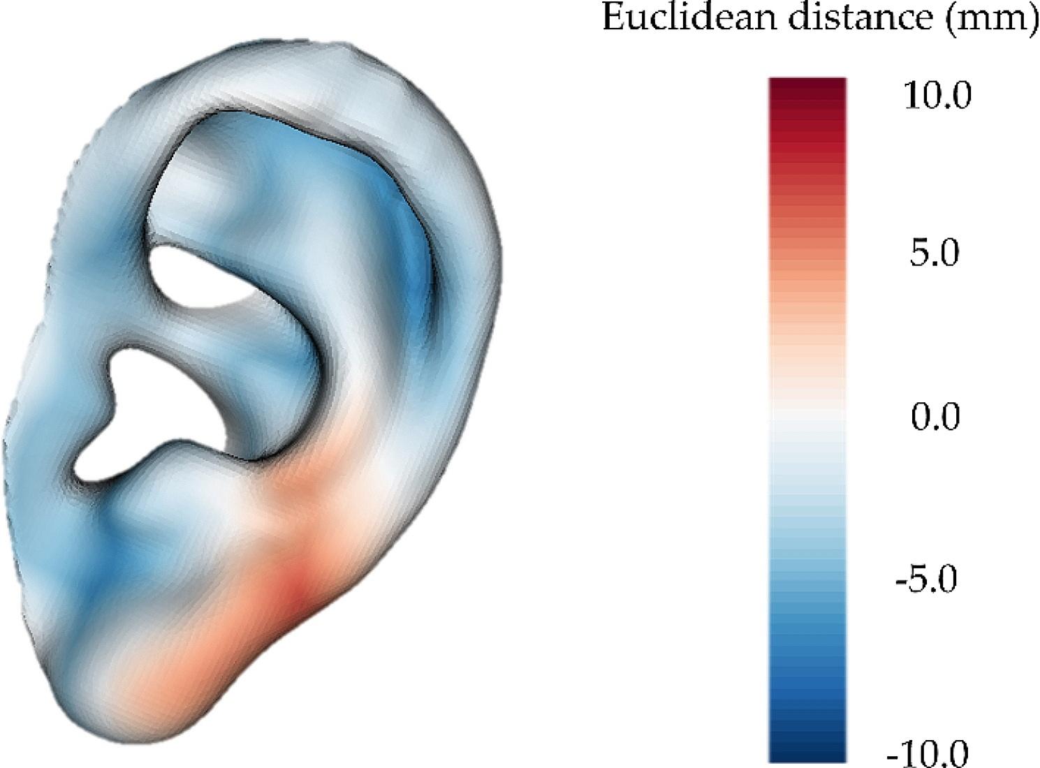
Results: Overall, the ears placed in the simulation scenario had a mean absolute deviation of 2.2 ± 1.7 mm with respect to the reference plan. During the surgical intervention, the reconstructed ear was 3.1 mm longer and 1.3 mm wider with respect to the ideal plan and had a positioning error of 2.7 ± 2.4 mm relative to the contralateral side. Note that in this case, additional morphometric variations were induced from inflammation and other issues intended to be addressed in a subsequent stage of surgery, which are independent of our proposed solution.
Conclusions: In this work we propose an innovative workflow that combines 3D printing and AR to improve ear reconstruction and positioning in microtia correction procedures. Our implementation in the surgical workflow showed good accuracy, empowering surgeons to attain consistent and objective outcomes.


 分享
分享

 求助内容:
求助内容: 应助结果提醒方式:
 扫码关注我们
