Christopher Gibson, Shirley C Wang, Arcturus Phoon, Nayana Thalanki Anantha, Kathryn Ottolino-Perry, Stephen Petropoulos, Zuha Qureshi, Vasanth Subramanian, Anam Shahid, Cristiana O'Brien, Steven Carcone, Suzanne Chung, Teresa Tsui, Viktor Son, Mayleen Sukhram, Fannong Meng, Susan J Done, Alexandra M Easson, Tulin Cil, Michael Reedijk, Wey L Leong, Ralph S DaCosta
{"title":"A handheld device for intra-cavity and ex vivo fluorescence imaging of breast conserving surgery margins with 5-aminolevulinic acid.","authors":"Christopher Gibson, Shirley C Wang, Arcturus Phoon, Nayana Thalanki Anantha, Kathryn Ottolino-Perry, Stephen Petropoulos, Zuha Qureshi, Vasanth Subramanian, Anam Shahid, Cristiana O'Brien, Steven Carcone, Suzanne Chung, Teresa Tsui, Viktor Son, Mayleen Sukhram, Fannong Meng, Susan J Done, Alexandra M Easson, Tulin Cil, Michael Reedijk, Wey L Leong, Ralph S DaCosta","doi":"10.1186/s42490-024-00079-9","DOIUrl":null,"url":null,"abstract":"<p><strong>Background: </strong>Visualization of cancer during breast conserving surgery (BCS) remains challenging; the BCS reoperation rate is reported to be 20-70% of patients. An urgent clinical need exists for real-time intraoperative visualization of breast carcinomas during BCS. We previously demonstrated the ability of a prototype imaging device to identify breast carcinoma in excised surgical specimens following 5-aminolevulinic acid (5-ALA) administration. However, this prototype device was not designed to image the surgical cavity for remaining carcinoma after the excised lumpectomy specimen is removed. A new handheld fluorescence (FL) imaging prototype device, designed to image both excised specimens and within the surgical cavity, was assessed in a clinical trial to evaluate its clinical utility for first-in-human, real-time intraoperative imaging during index BCS.</p><p><strong>Results: </strong>The imaging device combines consumer-grade imaging sensory technology with miniature light-emitting diodes (LEDs) and multiband optical filtering to capture high-resolution white light (WL) and FL digital images and videos. The technology allows for visualization of protoporphyrin IX (PpIX), which fluoresces red when excited by violet-blue light. To date, <math><mrow><mi>n</mi> <mo>=</mo> <mn>17</mn></mrow> </math> patients have received <math><mrow><mn>20</mn> <mfrac><mtext>mg</mtext> <mtext>kg</mtext></mfrac> </mrow> </math> bodyweight (BW) 5-ALA orally 2-4 h before imaging to facilitate the accumulation of PpIX within tumour cells. Tissue types were identified based on their colour appearance. Breast tumours in sectioned lumpectomies appeared red, which contrasted against the green connective tissues and orange-brown adipose tissues. In addition, ductal carcinoma in situ (DCIS) that was missed during intraoperative standard of care was identified at the surgical margin at <1 mm depth. In addition, artifacts due to the surgical drape, illumination, and blood within the surgical cavity were discovered.</p><p><strong>Conclusions: </strong>This study has demonstrated the detection of a grossly occult positive margin intraoperatively. Artifacts from imaging within the surgical cavity have been identified, and potential mitigations have been proposed.</p><p><strong>Trial registration: </strong>ClinicalTrials.gov Identifier: NCT01837225 (Trial start date is September 2010. It was registered to ClinicalTrials.gov retrospectively on April 23, 2013, then later updated on April 9, 2020, to reflect the introduction of the new imaging device.).</p>","PeriodicalId":72425,"journal":{"name":"BMC biomedical engineering","volume":"6 1","pages":"5"},"PeriodicalIF":0.0000,"publicationDate":"2024-06-01","publicationTypes":"Journal Article","fieldsOfStudy":null,"isOpenAccess":false,"openAccessPdf":"https://www.ncbi.nlm.nih.gov/pmc/articles/PMC11143723/pdf/","citationCount":"0","resultStr":null,"platform":"Semanticscholar","paperid":null,"PeriodicalName":"BMC biomedical engineering","FirstCategoryId":"1085","ListUrlMain":"https://doi.org/10.1186/s42490-024-00079-9","RegionNum":0,"RegionCategory":null,"ArticlePicture":[],"TitleCN":null,"AbstractTextCN":null,"PMCID":null,"EPubDate":"","PubModel":"","JCR":"","JCRName":"","Score":null,"Total":0}
引用次数: 0
Abstract
Background: Visualization of cancer during breast conserving surgery (BCS) remains challenging; the BCS reoperation rate is reported to be 20-70% of patients. An urgent clinical need exists for real-time intraoperative visualization of breast carcinomas during BCS. We previously demonstrated the ability of a prototype imaging device to identify breast carcinoma in excised surgical specimens following 5-aminolevulinic acid (5-ALA) administration. However, this prototype device was not designed to image the surgical cavity for remaining carcinoma after the excised lumpectomy specimen is removed. A new handheld fluorescence (FL) imaging prototype device, designed to image both excised specimens and within the surgical cavity, was assessed in a clinical trial to evaluate its clinical utility for first-in-human, real-time intraoperative imaging during index BCS.
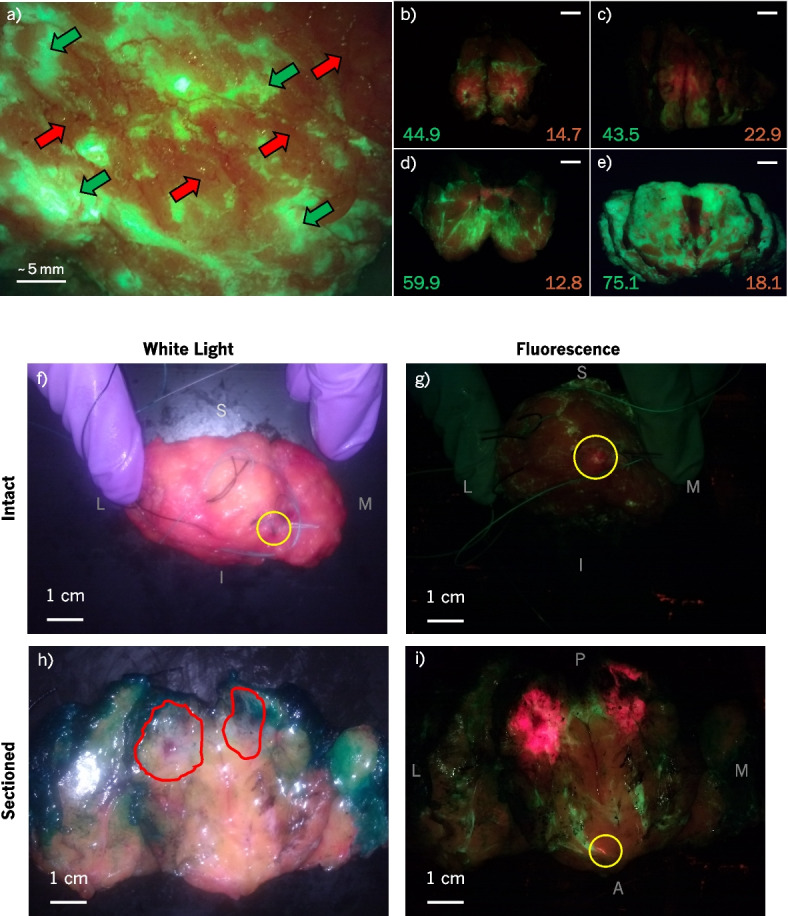
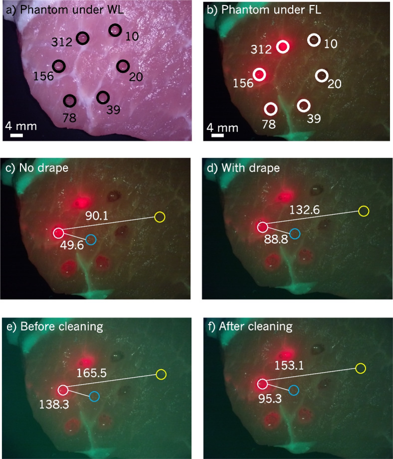
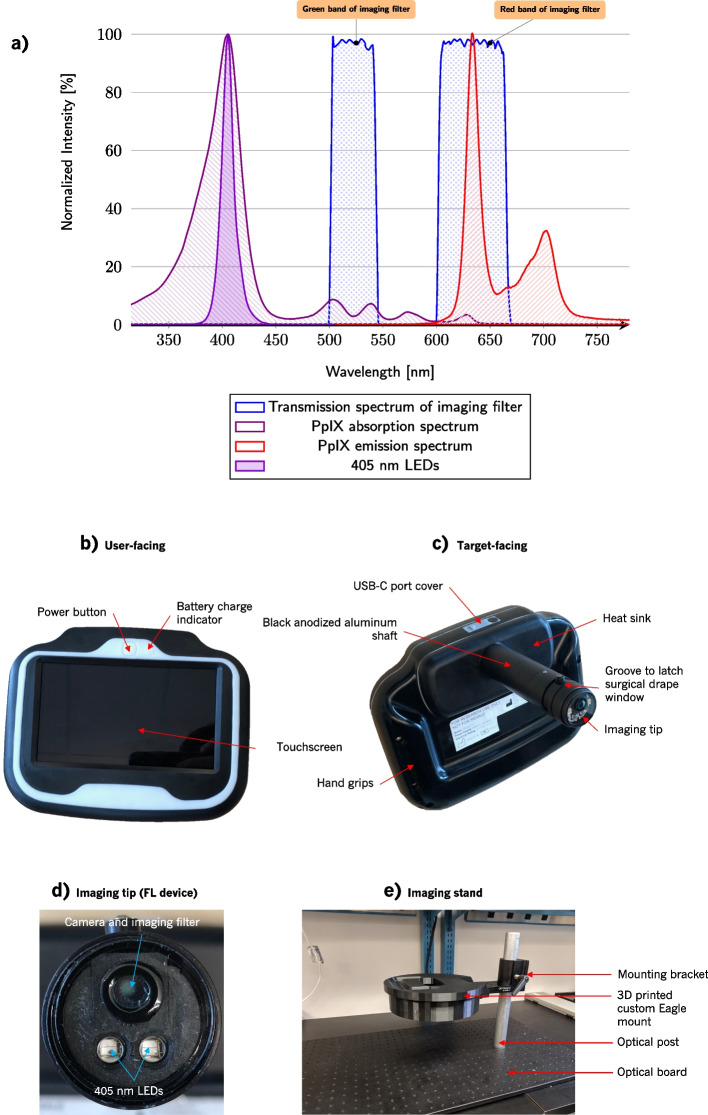
Results: The imaging device combines consumer-grade imaging sensory technology with miniature light-emitting diodes (LEDs) and multiband optical filtering to capture high-resolution white light (WL) and FL digital images and videos. The technology allows for visualization of protoporphyrin IX (PpIX), which fluoresces red when excited by violet-blue light. To date, patients have received bodyweight (BW) 5-ALA orally 2-4 h before imaging to facilitate the accumulation of PpIX within tumour cells. Tissue types were identified based on their colour appearance. Breast tumours in sectioned lumpectomies appeared red, which contrasted against the green connective tissues and orange-brown adipose tissues. In addition, ductal carcinoma in situ (DCIS) that was missed during intraoperative standard of care was identified at the surgical margin at <1 mm depth. In addition, artifacts due to the surgical drape, illumination, and blood within the surgical cavity were discovered.
Conclusions: This study has demonstrated the detection of a grossly occult positive margin intraoperatively. Artifacts from imaging within the surgical cavity have been identified, and potential mitigations have been proposed.
Trial registration: ClinicalTrials.gov Identifier: NCT01837225 (Trial start date is September 2010. It was registered to ClinicalTrials.gov retrospectively on April 23, 2013, then later updated on April 9, 2020, to reflect the introduction of the new imaging device.).


 分享
分享

 求助内容:
求助内容: 应助结果提醒方式:
 扫码关注我们
