{"title":"CIC::NUTM1 sarcomas occurred in soft tissues of upper limbs : a rare case report and literature review.","authors":"Lina Zhao, Huihua He, Jiacai Ren, Yabing Huang, Honglin Yan, Jingping Yuan","doi":"10.1186/s13000-024-01499-w","DOIUrl":null,"url":null,"abstract":"<p><strong>Background: </strong>CIC-rearranged sarcomas (CRS) represent a new entity of undifferentiated small round cell sarcoma belonging to the Ewing-like sarcomas family. CRS are the most common type. Fusion partners for the CIC gene include DUX4, FOXO4, and the recently recognizedNUTM1. Rare cases of CIC::NUTM1 sarcoma in pediatric patients have recently been reported in brain, kidney, bone, and soft tissues. However, such cases have not been identified in the soft tissues of the limbs.</p><p><strong>Case presentation: </strong>We reported a case of CIC::NUTM1 sarcoma located in the right upper limb of an 18-year-old man. The tumor displayed morphologic features typical of CIC::DUX4 sarcomas, with small- to medium-sized round cells, a lobular pattern, focal spindling, myxoid stroma, and patchy necrosis. The tumor diffusely expressed NUTM1, was positive for WT1cter at weak to moderate intensity, and was focally positive for CD99, while it was negative for keratins, EMA, P40, MyoD1, myogenin, NKX2.2, BCOR, and pan-TRK. Fluorescence in situ hybridization analyses revealed cleavage of the CIC and NUTM1 genes.</p><p><strong>Conclusion: </strong>CIC::NUTM1 sarcomas represent a novel molecular variant of CRS with a preference for the central nervous system and younger pediatric persons. Its morphology and phenotype may be mistaken for NUT carcinomas, and the behavior is more progressive than other forms of CRS. For this rare and newly discovered gene fusion variant, it is necessary to integrate molecular and immunohistochemical findings with morphologic features in the diagnosis of undifferentiated neoplasms.</p>","PeriodicalId":11237,"journal":{"name":"Diagnostic Pathology","volume":"19 1","pages":"76"},"PeriodicalIF":3.5000,"publicationDate":"2024-06-08","publicationTypes":"Journal Article","fieldsOfStudy":null,"isOpenAccess":false,"openAccessPdf":"https://www.ncbi.nlm.nih.gov/pmc/articles/PMC11162069/pdf/","citationCount":"0","resultStr":null,"platform":"Semanticscholar","paperid":null,"PeriodicalName":"Diagnostic Pathology","FirstCategoryId":"3","ListUrlMain":"https://doi.org/10.1186/s13000-024-01499-w","RegionNum":3,"RegionCategory":"医学","ArticlePicture":[],"TitleCN":null,"AbstractTextCN":null,"PMCID":null,"EPubDate":"","PubModel":"","JCR":"Q2","JCRName":"PATHOLOGY","Score":null,"Total":0}
引用次数: 0
Abstract
Background: CIC-rearranged sarcomas (CRS) represent a new entity of undifferentiated small round cell sarcoma belonging to the Ewing-like sarcomas family. CRS are the most common type. Fusion partners for the CIC gene include DUX4, FOXO4, and the recently recognizedNUTM1. Rare cases of CIC::NUTM1 sarcoma in pediatric patients have recently been reported in brain, kidney, bone, and soft tissues. However, such cases have not been identified in the soft tissues of the limbs.
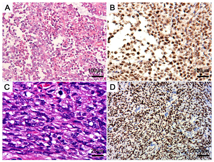
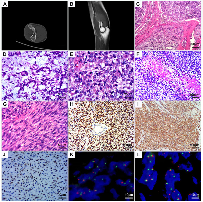
Case presentation: We reported a case of CIC::NUTM1 sarcoma located in the right upper limb of an 18-year-old man. The tumor displayed morphologic features typical of CIC::DUX4 sarcomas, with small- to medium-sized round cells, a lobular pattern, focal spindling, myxoid stroma, and patchy necrosis. The tumor diffusely expressed NUTM1, was positive for WT1cter at weak to moderate intensity, and was focally positive for CD99, while it was negative for keratins, EMA, P40, MyoD1, myogenin, NKX2.2, BCOR, and pan-TRK. Fluorescence in situ hybridization analyses revealed cleavage of the CIC and NUTM1 genes.
Conclusion: CIC::NUTM1 sarcomas represent a novel molecular variant of CRS with a preference for the central nervous system and younger pediatric persons. Its morphology and phenotype may be mistaken for NUT carcinomas, and the behavior is more progressive than other forms of CRS. For this rare and newly discovered gene fusion variant, it is necessary to integrate molecular and immunohistochemical findings with morphologic features in the diagnosis of undifferentiated neoplasms.
期刊介绍:
Diagnostic Pathology is an open access, peer-reviewed, online journal that considers research in surgical and clinical pathology, immunology, and biology, with a special focus on cutting-edge approaches in diagnostic pathology and tissue-based therapy. The journal covers all aspects of surgical pathology, including classic diagnostic pathology, prognosis-related diagnosis (tumor stages, prognosis markers, such as MIB-percentage, hormone receptors, etc.), and therapy-related findings. The journal also focuses on the technological aspects of pathology, including molecular biology techniques, morphometry aspects (stereology, DNA analysis, syntactic structure analysis), communication aspects (telecommunication, virtual microscopy, virtual pathology institutions, etc.), and electronic education and quality assurance (for example interactive publication, on-line references with automated updating, etc.).




 京公网安备 11010802042870号
京ICP备2023020795号-1
京公网安备 11010802042870号
京ICP备2023020795号-1

 分享
分享
 求助内容:
求助内容: 应助结果提醒方式:
 扫码关注我们
