Nityanand Jain, Liga Jaunozolina, Inga Putraima, Kaspars Auslands, Andrejs Millers
{"title":"Delayed spinal arachnoiditis with syringomyelia following aneurysmal subarachnoid haemorrhage: a case report with patient experience.","authors":"Nityanand Jain, Liga Jaunozolina, Inga Putraima, Kaspars Auslands, Andrejs Millers","doi":"10.1038/s41394-024-00654-1","DOIUrl":null,"url":null,"abstract":"<p><strong>Background and importance: </strong>Syringomyelia, or the formation of fluid-filled cysts within the spinal cord, associated with delayed spinal arachnoiditis is an uncommon complication of aneurysmal subarachnoid haemorrhage. To date, about 18 cases have been reported in medical literature, with just two reported in patients under the age of 35 years.</p><p><strong>Clinical presentation: </strong>A 27-year-old female patient complained of sudden, severe headaches in the occipital region, nuchal rigidity, and drowsiness when she presented at our institution. A head computed tomography scan revealed intraventricular bleeding in the lateral and fourth ventricles with more extensive haemorrhaging in the frontal horns. A left posterior inferior cerebellar artery (PICA) aneurysm was confirmed via digital subtraction angiogram, and endovascular embolization was done. Two years later, the patient reported intense pain in the lower back along with symptoms suggestive of spinal cord compression. Spinal magnetic resonance imaging (MRI) showed spinal adhesions from C1 to L4, syringomyelia with some vasogenic oedema extending from T3 to T9 level, and a cyst in the lumbar region. Consequently, a right hemilaminectomy was performed along with microsurgical release of arachnoid adhesions and placement of a subdural drain. Radiological and symptomatic improvements were observed. Since then, the patient's clinical condition has remained stable during the past three years of follow-up visits.</p><p><strong>Conclusions: </strong>Literature on optimal treatment modalities and patient prognosis is scarce and debated. The time for symptom improvement depends on the level and extent of spinal cord involvement. Rehabilitation may be required for most patients, as complete symptomatic recovery may not be attainable.</p>","PeriodicalId":22079,"journal":{"name":"Spinal Cord Series and Cases","volume":"10 1","pages":"41"},"PeriodicalIF":0.9000,"publicationDate":"2024-06-10","publicationTypes":"Journal Article","fieldsOfStudy":null,"isOpenAccess":false,"openAccessPdf":"https://www.ncbi.nlm.nih.gov/pmc/articles/PMC11165000/pdf/","citationCount":"0","resultStr":null,"platform":"Semanticscholar","paperid":null,"PeriodicalName":"Spinal Cord Series and Cases","FirstCategoryId":"1085","ListUrlMain":"https://doi.org/10.1038/s41394-024-00654-1","RegionNum":0,"RegionCategory":null,"ArticlePicture":[],"TitleCN":null,"AbstractTextCN":null,"PMCID":null,"EPubDate":"","PubModel":"","JCR":"Q4","JCRName":"CLINICAL NEUROLOGY","Score":null,"Total":0}
引用次数: 0
Abstract
Background and importance: Syringomyelia, or the formation of fluid-filled cysts within the spinal cord, associated with delayed spinal arachnoiditis is an uncommon complication of aneurysmal subarachnoid haemorrhage. To date, about 18 cases have been reported in medical literature, with just two reported in patients under the age of 35 years.
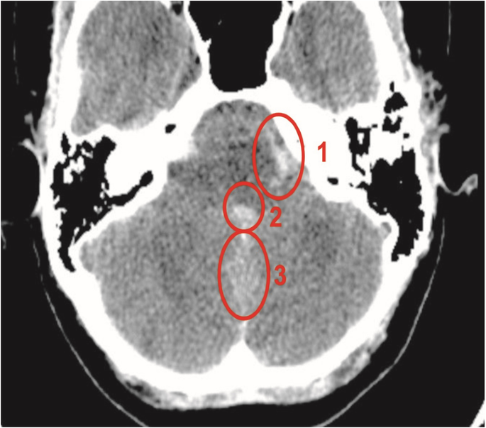
Clinical presentation: A 27-year-old female patient complained of sudden, severe headaches in the occipital region, nuchal rigidity, and drowsiness when she presented at our institution. A head computed tomography scan revealed intraventricular bleeding in the lateral and fourth ventricles with more extensive haemorrhaging in the frontal horns. A left posterior inferior cerebellar artery (PICA) aneurysm was confirmed via digital subtraction angiogram, and endovascular embolization was done. Two years later, the patient reported intense pain in the lower back along with symptoms suggestive of spinal cord compression. Spinal magnetic resonance imaging (MRI) showed spinal adhesions from C1 to L4, syringomyelia with some vasogenic oedema extending from T3 to T9 level, and a cyst in the lumbar region. Consequently, a right hemilaminectomy was performed along with microsurgical release of arachnoid adhesions and placement of a subdural drain. Radiological and symptomatic improvements were observed. Since then, the patient's clinical condition has remained stable during the past three years of follow-up visits.
Conclusions: Literature on optimal treatment modalities and patient prognosis is scarce and debated. The time for symptom improvement depends on the level and extent of spinal cord involvement. Rehabilitation may be required for most patients, as complete symptomatic recovery may not be attainable.


 分享
分享
 求助内容:
求助内容: 应助结果提醒方式:
 扫码关注我们
