Artificial intelligence-driven computer aided diagnosis system provides similar diagnosis value compared with doctors' evaluation in lung cancer screening.
{"title":"Artificial intelligence-driven computer aided diagnosis system provides similar diagnosis value compared with doctors' evaluation in lung cancer screening.","authors":"Shan Gao, Zexuan Xu, Wanli Kang, Xinna Lv, Naihui Chu, Shaofa Xu, Dailun Hou","doi":"10.1186/s12880-024-01288-3","DOIUrl":null,"url":null,"abstract":"<p><strong>Objective: </strong>To evaluate the consistency between doctors and artificial intelligence (AI) software in analysing and diagnosing pulmonary nodules, and assess whether the characteristics of pulmonary nodules derived from the two methods are consistent for the interpretation of carcinomatous nodules.</p><p><strong>Materials and methods: </strong>This retrospective study analysed participants aged 40-74 in the local area from 2011 to 2013. Pulmonary nodules were examined radiologically using a low-dose chest CT scan, evaluated by an expert panel of doctors in radiology, oncology, and thoracic departments, as well as a computer-aided diagnostic(CAD) system based on the three-dimensional(3D) convolutional neural network (CNN) with DenseNet architecture(InferRead CT Lung, IRCL). Consistency tests were employed to assess the uniformity of the radiological characteristics of the pulmonary nodules. The receiver operating characteristic (ROC) curve was used to evaluate the diagnostic accuracy. Logistic regression analysis is utilized to determine whether the two methods yield the same predictive factors for cancerous nodules.</p><p><strong>Results: </strong>A total of 570 subjects were included in this retrospective study. The AI software demonstrated high consistency with the panel's evaluation in determining the position and diameter of the pulmonary nodules (kappa = 0.883, concordance correlation coefficient (CCC) = 0.809, p = 0.000). The comparison of the solid nodules' attenuation characteristics also showed acceptable consistency (kappa = 0.503). In patients diagnosed with lung cancer, the area under the curve (AUC) for the panel and AI were 0.873 (95%CI: 0.829-0.909) and 0.921 (95%CI: 0.884-0.949), respectively. However, there was no significant difference (p = 0.0950). The maximum diameter, solid nodules, subsolid nodules were the crucial factors for interpreting carcinomatous nodules in the analysis of expert panel and IRCL pulmonary nodule characteristics.</p><p><strong>Conclusion: </strong>AI software can assist doctors in diagnosing nodules and is consistent with doctors' evaluations and diagnosis of pulmonary nodules.</p>","PeriodicalId":9020,"journal":{"name":"BMC Medical Imaging","volume":"24 1","pages":"141"},"PeriodicalIF":3.2000,"publicationDate":"2024-06-11","publicationTypes":"Journal Article","fieldsOfStudy":null,"isOpenAccess":false,"openAccessPdf":"https://www.ncbi.nlm.nih.gov/pmc/articles/PMC11165751/pdf/","citationCount":"0","resultStr":null,"platform":"Semanticscholar","paperid":null,"PeriodicalName":"BMC Medical Imaging","FirstCategoryId":"3","ListUrlMain":"https://doi.org/10.1186/s12880-024-01288-3","RegionNum":3,"RegionCategory":"医学","ArticlePicture":[],"TitleCN":null,"AbstractTextCN":null,"PMCID":null,"EPubDate":"","PubModel":"","JCR":"Q2","JCRName":"RADIOLOGY, NUCLEAR MEDICINE & MEDICAL IMAGING","Score":null,"Total":0}
引用次数: 0
Abstract
Objective: To evaluate the consistency between doctors and artificial intelligence (AI) software in analysing and diagnosing pulmonary nodules, and assess whether the characteristics of pulmonary nodules derived from the two methods are consistent for the interpretation of carcinomatous nodules.
Materials and methods: This retrospective study analysed participants aged 40-74 in the local area from 2011 to 2013. Pulmonary nodules were examined radiologically using a low-dose chest CT scan, evaluated by an expert panel of doctors in radiology, oncology, and thoracic departments, as well as a computer-aided diagnostic(CAD) system based on the three-dimensional(3D) convolutional neural network (CNN) with DenseNet architecture(InferRead CT Lung, IRCL). Consistency tests were employed to assess the uniformity of the radiological characteristics of the pulmonary nodules. The receiver operating characteristic (ROC) curve was used to evaluate the diagnostic accuracy. Logistic regression analysis is utilized to determine whether the two methods yield the same predictive factors for cancerous nodules.
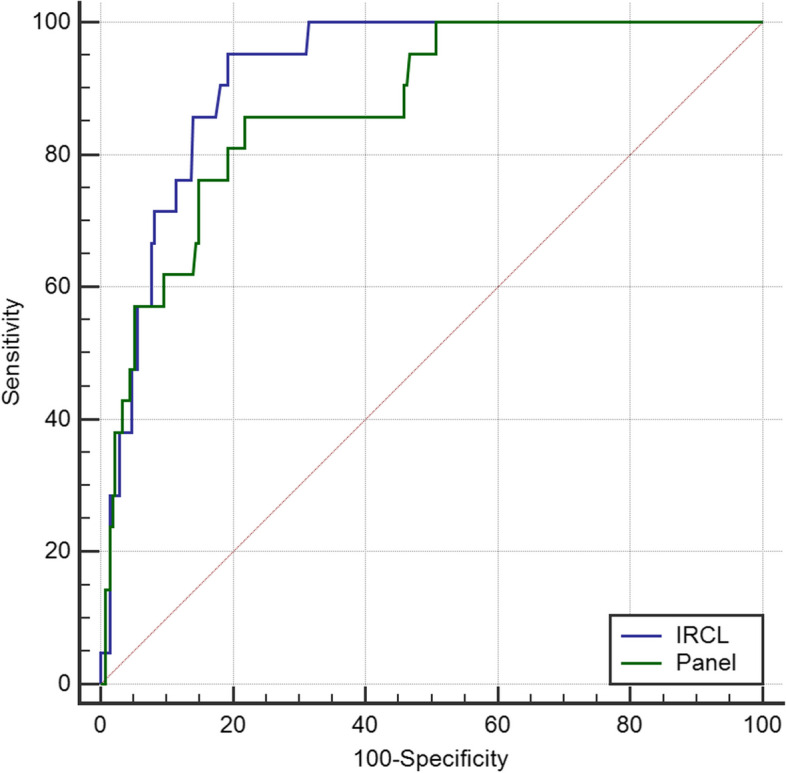
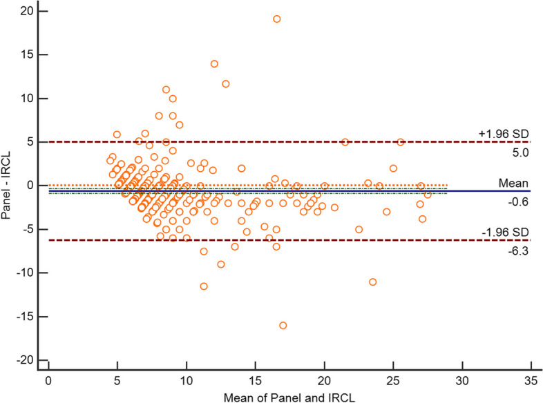
Results: A total of 570 subjects were included in this retrospective study. The AI software demonstrated high consistency with the panel's evaluation in determining the position and diameter of the pulmonary nodules (kappa = 0.883, concordance correlation coefficient (CCC) = 0.809, p = 0.000). The comparison of the solid nodules' attenuation characteristics also showed acceptable consistency (kappa = 0.503). In patients diagnosed with lung cancer, the area under the curve (AUC) for the panel and AI were 0.873 (95%CI: 0.829-0.909) and 0.921 (95%CI: 0.884-0.949), respectively. However, there was no significant difference (p = 0.0950). The maximum diameter, solid nodules, subsolid nodules were the crucial factors for interpreting carcinomatous nodules in the analysis of expert panel and IRCL pulmonary nodule characteristics.
Conclusion: AI software can assist doctors in diagnosing nodules and is consistent with doctors' evaluations and diagnosis of pulmonary nodules.
期刊介绍:
BMC Medical Imaging is an open access journal publishing original peer-reviewed research articles in the development, evaluation, and use of imaging techniques and image processing tools to diagnose and manage disease.


 分享
分享
 求助内容:
求助内容: 应助结果提醒方式:
 扫码关注我们
