{"title":"Developmental morpho-analysis of the caecum in Japanese quail embryos (Coturnix coturnix japonica)","authors":"Fatma Abdelhakeem","doi":"10.1002/jemt.24632","DOIUrl":null,"url":null,"abstract":"<div>\n \n \n <section>\n \n <p>In the current study, we are focusing on the microanatomical structure of quail caecum during the prehatching time to try to understand the function and the role of each cell-built quail caecum reaching how caecum plays an essential role in immunity and absorption. The morpho-developmental features of the quail caecum were described in detail daily from the third incubation day (ID) till hatching time, investigating the gross morphology, microscopic, and ultrastructure using light and scanning electron microscope. The embryonic caecum appeared grossly as two lateral outpocketings with blinded ends, emerging laterally at the junction between the small and large intestine (the ileocaecal junction). The primordia of two caeca, represented by two lateral swellings from the hindgut on the fourth ID, continued growing till the day of hatching, where the caecal wall consisted of three apparent layers: mucosa, musculosa, and serosa. At the time of hatching, the quail caecum was still not fully mature and will continue growing posthatching. The findings in this study can be applied in further studies intended to understand the physiological mechanisms of the caecum during prehatching and posthatching periods.</p>\n </section>\n \n <section>\n \n <h3> Research Highlights</h3>\n \n <div>\n <ul>\n \n <li>Caecum is one of the hindgut derivatives that started as two lateral swellings.</li>\n \n <li>The caecal wall consisted of three layers; mucosa, musculosa, and serosa.</li>\n \n <li>The caecum plays an essential role in immunity maintenance.</li>\n \n <li>Caecum continues to grow posthatching as it is not fully mature at hatching time.</li>\n </ul>\n </div>\n </section>\n </div>","PeriodicalId":18684,"journal":{"name":"Microscopy Research and Technique","volume":"87 11","pages":"2540-2554"},"PeriodicalIF":2.1000,"publicationDate":"2024-06-12","publicationTypes":"Journal Article","fieldsOfStudy":null,"isOpenAccess":false,"openAccessPdf":"","citationCount":"0","resultStr":null,"platform":"Semanticscholar","paperid":null,"PeriodicalName":"Microscopy Research and Technique","FirstCategoryId":"5","ListUrlMain":"https://analyticalsciencejournals.onlinelibrary.wiley.com/doi/10.1002/jemt.24632","RegionNum":3,"RegionCategory":"工程技术","ArticlePicture":[],"TitleCN":null,"AbstractTextCN":null,"PMCID":null,"EPubDate":"","PubModel":"","JCR":"Q2","JCRName":"ANATOMY & MORPHOLOGY","Score":null,"Total":0}
引用次数: 0
Abstract
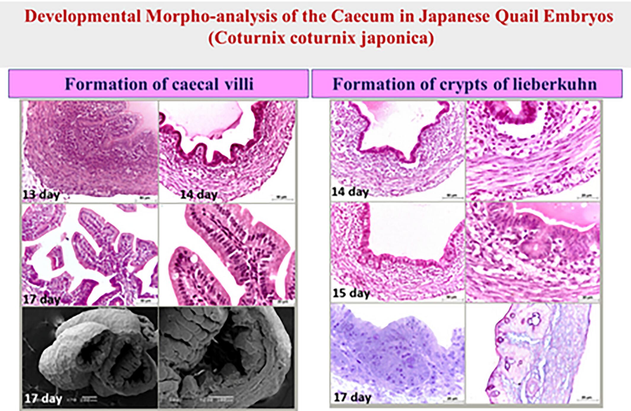
In the current study, we are focusing on the microanatomical structure of quail caecum during the prehatching time to try to understand the function and the role of each cell-built quail caecum reaching how caecum plays an essential role in immunity and absorption. The morpho-developmental features of the quail caecum were described in detail daily from the third incubation day (ID) till hatching time, investigating the gross morphology, microscopic, and ultrastructure using light and scanning electron microscope. The embryonic caecum appeared grossly as two lateral outpocketings with blinded ends, emerging laterally at the junction between the small and large intestine (the ileocaecal junction). The primordia of two caeca, represented by two lateral swellings from the hindgut on the fourth ID, continued growing till the day of hatching, where the caecal wall consisted of three apparent layers: mucosa, musculosa, and serosa. At the time of hatching, the quail caecum was still not fully mature and will continue growing posthatching. The findings in this study can be applied in further studies intended to understand the physiological mechanisms of the caecum during prehatching and posthatching periods.
Research Highlights
Caecum is one of the hindgut derivatives that started as two lateral swellings.
The caecal wall consisted of three layers; mucosa, musculosa, and serosa.
The caecum plays an essential role in immunity maintenance.
Caecum continues to grow posthatching as it is not fully mature at hatching time.
期刊介绍:
Microscopy Research and Technique (MRT) publishes articles on all aspects of advanced microscopy original architecture and methodologies with applications in the biological, clinical, chemical, and materials sciences. Original basic and applied research as well as technical papers dealing with the various subsets of microscopy are encouraged. MRT is the right form for those developing new microscopy methods or using the microscope to answer key questions in basic and applied research.


 分享
分享
 求助内容:
求助内容: 应助结果提醒方式:
 扫码关注我们
