Association between medial meniscal extrusion and knee structural progression in adults with symptomatic knee osteoarthritis - a prospective cohort study.
Mengjie Zeng, Flavia M Cicuttini, Anita E Wluka, Graeme Jones, Catherine L Hill, Changhai Ding, Yuanyuan Wang
{"title":"Association between medial meniscal extrusion and knee structural progression in adults with symptomatic knee osteoarthritis - a prospective cohort study.","authors":"Mengjie Zeng, Flavia M Cicuttini, Anita E Wluka, Graeme Jones, Catherine L Hill, Changhai Ding, Yuanyuan Wang","doi":"10.1007/s00256-024-04731-2","DOIUrl":null,"url":null,"abstract":"<p><strong>Objective: </strong>To examine the association between medial meniscal extrusion and structural progression in adults with symptomatic knee osteoarthritis (OA).</p><p><strong>Methods: </strong>This prospective cohort study examined 176 participants with symptomatic knee OA recruited into a randomised controlled trial. The participants underwent magnetic resonance imaging (MRI) of the study knee at baseline and approximately 2 years later. Meniscal extrusion, tibial cartilage volume, and tibiofemoral bone marrow lesions (BMLs) were measured from MRI using validated methods.</p><p><strong>Results: </strong>Participants with medial meniscal extrusion ≥ 3 mm had a higher prevalence of lateral tibiofemoral BMLs at baseline (OR = 2.21, 95% CI 1.06-4.61, p = 0.035), and those with medial meniscal extrusion 2-3 mm had a higher likelihood of lateral BML worsening over 2 years (OR = 3.76, 95% CI 1.35-10.52, p = 0.011), compared with those with medial meniscal extrusion < 2 mm. Participants with stable medial meniscal extrusion had a lower likelihood of lateral BML worsening compared with those with regression of medial meniscal extrusion over 2 years (OR = 0.20, 95% CI 0.07-0.56, p = 0.002). There were no associations between medial meniscal extrusion and tibial cartilage volume or medial tibiofemoral BMLs.</p><p><strong>Conclusions: </strong>Our study showed associations between medial meniscal extrusion and baseline prevalence and worsening over 2 years of lateral tibiofemoral BMLs in people with symptomatic knee OA. Although the reasons for the lack of associations in the medial compartment are not clear, our results suggest a role of medial meniscal extrusion in predicting structural progression in lateral knee OA and that meniscal extrusion might be a potential target in the management of knee OA.</p>","PeriodicalId":21783,"journal":{"name":"Skeletal Radiology","volume":" ","pages":"219-228"},"PeriodicalIF":2.2000,"publicationDate":"2025-02-01","publicationTypes":"Journal Article","fieldsOfStudy":null,"isOpenAccess":false,"openAccessPdf":"https://www.ncbi.nlm.nih.gov/pmc/articles/PMC11652669/pdf/","citationCount":"0","resultStr":null,"platform":"Semanticscholar","paperid":null,"PeriodicalName":"Skeletal Radiology","FirstCategoryId":"3","ListUrlMain":"https://doi.org/10.1007/s00256-024-04731-2","RegionNum":3,"RegionCategory":"医学","ArticlePicture":[],"TitleCN":null,"AbstractTextCN":null,"PMCID":null,"EPubDate":"2024/6/15 0:00:00","PubModel":"Epub","JCR":"Q2","JCRName":"ORTHOPEDICS","Score":null,"Total":0}
引用次数: 0
Abstract
Objective: To examine the association between medial meniscal extrusion and structural progression in adults with symptomatic knee osteoarthritis (OA).
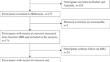
Methods: This prospective cohort study examined 176 participants with symptomatic knee OA recruited into a randomised controlled trial. The participants underwent magnetic resonance imaging (MRI) of the study knee at baseline and approximately 2 years later. Meniscal extrusion, tibial cartilage volume, and tibiofemoral bone marrow lesions (BMLs) were measured from MRI using validated methods.
Results: Participants with medial meniscal extrusion ≥ 3 mm had a higher prevalence of lateral tibiofemoral BMLs at baseline (OR = 2.21, 95% CI 1.06-4.61, p = 0.035), and those with medial meniscal extrusion 2-3 mm had a higher likelihood of lateral BML worsening over 2 years (OR = 3.76, 95% CI 1.35-10.52, p = 0.011), compared with those with medial meniscal extrusion < 2 mm. Participants with stable medial meniscal extrusion had a lower likelihood of lateral BML worsening compared with those with regression of medial meniscal extrusion over 2 years (OR = 0.20, 95% CI 0.07-0.56, p = 0.002). There were no associations between medial meniscal extrusion and tibial cartilage volume or medial tibiofemoral BMLs.
Conclusions: Our study showed associations between medial meniscal extrusion and baseline prevalence and worsening over 2 years of lateral tibiofemoral BMLs in people with symptomatic knee OA. Although the reasons for the lack of associations in the medial compartment are not clear, our results suggest a role of medial meniscal extrusion in predicting structural progression in lateral knee OA and that meniscal extrusion might be a potential target in the management of knee OA.
期刊介绍:
Skeletal Radiology provides a forum for the dissemination of current knowledge and information dealing with disorders of the musculoskeletal system including the spine. While emphasizing the radiological aspects of the many varied skeletal abnormalities, the journal also adopts an interdisciplinary approach, reflecting the membership of the International Skeletal Society. Thus, the anatomical, pathological, physiological, clinical, metabolic and epidemiological aspects of the many entities affecting the skeleton receive appropriate consideration.
This is the Journal of the International Skeletal Society and the Official Journal of the Society of Skeletal Radiology and the Australasian Musculoskelelal Imaging Group.


 分享
分享
 求助内容:
求助内容: 应助结果提醒方式:
 扫码关注我们
