Kenan Morani, Esra Kaya Ayana, Dimitrios Kollias, Devrim Unay
{"title":"COVID-19 Detection from Computed Tomography Images Using Slice Processing Techniques and a Modified Xception Classifier.","authors":"Kenan Morani, Esra Kaya Ayana, Dimitrios Kollias, Devrim Unay","doi":"10.1155/2024/9962839","DOIUrl":null,"url":null,"abstract":"<p><p>This paper extends our previous method for COVID-19 diagnosis, proposing an enhanced solution for detecting COVID-19 from computed tomography (CT) images using a lean transfer learning-based model. To decrease model misclassifications, two key steps of image processing were employed. Firstly, the uppermost and lowermost slices were removed, preserving sixty percent of each patient's slices. Secondly, all slices underwent manual cropping to emphasize the lung areas. Subsequently, resized CT scans (224 × 224) were input into an Xception transfer learning model with a modified output. Both Xception's architecture and pretrained weights were leveraged in the method. A big and rigorously annotated database of CT images was used to verify the method. The number of patients/subjects in the dataset is more than 5000, and the number and shape of the slices in each CT scan varies greatly. Verification was made both on the validation partition and on the test partition of unseen images. Results on the COV19-CT database showcased not only improvement from our previous solution and the baseline but also comparable performance to the highest-achieving methods on the same dataset. Further validation studies could explore the scalability and adaptability of the developed methodologies across diverse healthcare settings and patient populations. Additionally, investigating the integration of advanced image processing techniques, such as automated region of interest detection and segmentation algorithms, could enhance the efficiency and accuracy of COVID-19 diagnosis.</p>","PeriodicalId":47063,"journal":{"name":"International Journal of Biomedical Imaging","volume":"2024 ","pages":"9962839"},"PeriodicalIF":1.3000,"publicationDate":"2024-05-24","publicationTypes":"Journal Article","fieldsOfStudy":null,"isOpenAccess":false,"openAccessPdf":"https://www.ncbi.nlm.nih.gov/pmc/articles/PMC11178392/pdf/","citationCount":"0","resultStr":null,"platform":"Semanticscholar","paperid":null,"PeriodicalName":"International Journal of Biomedical Imaging","FirstCategoryId":"1085","ListUrlMain":"https://doi.org/10.1155/2024/9962839","RegionNum":0,"RegionCategory":null,"ArticlePicture":[],"TitleCN":null,"AbstractTextCN":null,"PMCID":null,"EPubDate":"2024/1/1 0:00:00","PubModel":"eCollection","JCR":"Q2","JCRName":"ENGINEERING, BIOMEDICAL","Score":null,"Total":0}
引用次数: 0
Abstract
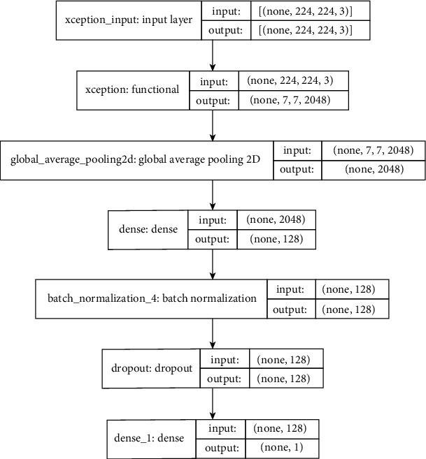
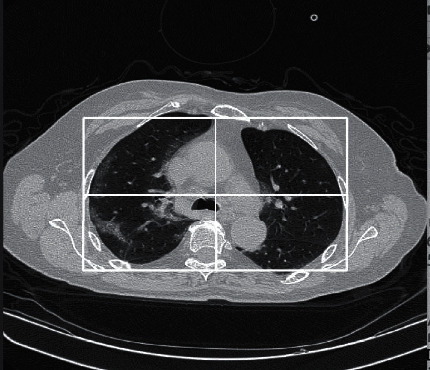
This paper extends our previous method for COVID-19 diagnosis, proposing an enhanced solution for detecting COVID-19 from computed tomography (CT) images using a lean transfer learning-based model. To decrease model misclassifications, two key steps of image processing were employed. Firstly, the uppermost and lowermost slices were removed, preserving sixty percent of each patient's slices. Secondly, all slices underwent manual cropping to emphasize the lung areas. Subsequently, resized CT scans (224 × 224) were input into an Xception transfer learning model with a modified output. Both Xception's architecture and pretrained weights were leveraged in the method. A big and rigorously annotated database of CT images was used to verify the method. The number of patients/subjects in the dataset is more than 5000, and the number and shape of the slices in each CT scan varies greatly. Verification was made both on the validation partition and on the test partition of unseen images. Results on the COV19-CT database showcased not only improvement from our previous solution and the baseline but also comparable performance to the highest-achieving methods on the same dataset. Further validation studies could explore the scalability and adaptability of the developed methodologies across diverse healthcare settings and patient populations. Additionally, investigating the integration of advanced image processing techniques, such as automated region of interest detection and segmentation algorithms, could enhance the efficiency and accuracy of COVID-19 diagnosis.
期刊介绍:
The International Journal of Biomedical Imaging is managed by a board of editors comprising internationally renowned active researchers. The journal is freely accessible online and also offered for purchase in print format. It employs a web-based review system to ensure swift turnaround times while maintaining high standards. In addition to regular issues, special issues are organized by guest editors. The subject areas covered include (but are not limited to):
Digital radiography and tomosynthesis
X-ray computed tomography (CT)
Magnetic resonance imaging (MRI)
Single photon emission computed tomography (SPECT)
Positron emission tomography (PET)
Ultrasound imaging
Diffuse optical tomography, coherence, fluorescence, bioluminescence tomography, impedance tomography
Neutron imaging for biomedical applications
Magnetic and optical spectroscopy, and optical biopsy
Optical, electron, scanning tunneling/atomic force microscopy
Small animal imaging
Functional, cellular, and molecular imaging
Imaging assays for screening and molecular analysis
Microarray image analysis and bioinformatics
Emerging biomedical imaging techniques
Imaging modality fusion
Biomedical imaging instrumentation
Biomedical image processing, pattern recognition, and analysis
Biomedical image visualization, compression, transmission, and storage
Imaging and modeling related to systems biology and systems biomedicine
Applied mathematics, applied physics, and chemistry related to biomedical imaging
Grid-enabling technology for biomedical imaging and informatics


 分享
分享
 求助内容:
求助内容: 应助结果提醒方式:
 扫码关注我们
