{"title":"7T MRI in cerebrovascular disorders: From large artery abnormalities to small vessel disease","authors":"","doi":"10.1016/j.metrad.2024.100085","DOIUrl":null,"url":null,"abstract":"<div><p>There is a growing interest and adoption of 7 Tesla (T) magnetic resonance imaging (MRI) in the field of medicine and research. In the domain of neuroimaging, 7T MRI shows notable advantages over lower field strength MRI systems by offering improved visualization of anatomical structures, enhanced lesion conspicuity, and better characterization of pathological processes. Cerebrovascular disease, which involves a spectrum of etiologies from large artery abnormalities to small vessel disease, is a leading cause of morbidity and mortality worldwide. Imaging plays an indispensable role in the diagnosis and treatment of cerebrovascular diseases. The excellence in imaging capabilities of 7T MRI can achieve multi-scale, high-precision imaging requirements from large artery disease assessment to small vessel disease assessment, which presents a variety of clinical applications and significant potential for clinical transformation. In this review, we firstly reviewed the literature focusing on technique aspects, comparing 7T with the clinically well-established 3T and 1.5T MRI systems. Then, we reviewed published studies to showcase the state-of-the-art progress in the assessment of cerebrovascular disease at 7T. Additionally, we discussed the challenges and perspectives of 7T techniques.</p></div>","PeriodicalId":100921,"journal":{"name":"Meta-Radiology","volume":"2 3","pages":"Article 100085"},"PeriodicalIF":0.0000,"publicationDate":"2024-09-01","publicationTypes":"Journal Article","fieldsOfStudy":null,"isOpenAccess":false,"openAccessPdf":"https://www.sciencedirect.com/science/article/pii/S2950162824000389/pdfft?md5=4fa5a90f11cc6834a872f3bb8b907d5f&pid=1-s2.0-S2950162824000389-main.pdf","citationCount":"0","resultStr":null,"platform":"Semanticscholar","paperid":null,"PeriodicalName":"Meta-Radiology","FirstCategoryId":"1085","ListUrlMain":"https://www.sciencedirect.com/science/article/pii/S2950162824000389","RegionNum":0,"RegionCategory":null,"ArticlePicture":[],"TitleCN":null,"AbstractTextCN":null,"PMCID":null,"EPubDate":"2024/6/12 0:00:00","PubModel":"Epub","JCR":"","JCRName":"","Score":null,"Total":0}
引用次数: 0
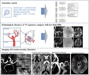
Abstract
There is a growing interest and adoption of 7 Tesla (T) magnetic resonance imaging (MRI) in the field of medicine and research. In the domain of neuroimaging, 7T MRI shows notable advantages over lower field strength MRI systems by offering improved visualization of anatomical structures, enhanced lesion conspicuity, and better characterization of pathological processes. Cerebrovascular disease, which involves a spectrum of etiologies from large artery abnormalities to small vessel disease, is a leading cause of morbidity and mortality worldwide. Imaging plays an indispensable role in the diagnosis and treatment of cerebrovascular diseases. The excellence in imaging capabilities of 7T MRI can achieve multi-scale, high-precision imaging requirements from large artery disease assessment to small vessel disease assessment, which presents a variety of clinical applications and significant potential for clinical transformation. In this review, we firstly reviewed the literature focusing on technique aspects, comparing 7T with the clinically well-established 3T and 1.5T MRI systems. Then, we reviewed published studies to showcase the state-of-the-art progress in the assessment of cerebrovascular disease at 7T. Additionally, we discussed the challenges and perspectives of 7T techniques.


 分享
分享

 求助内容:
求助内容: 应助结果提醒方式:
 扫码关注我们
