Tommaso Bo, Elia Pascucci, Simone Capuani, Jocelyn Nikita Campa-Carranza, Letizia Franco, Marco Farina, Jacopo Secco, Sara Becchi, Rosanna Cavazzana, Ashley L. Joubert, Nathanael Hernandez, Corrine Ying Xuan Chua, Alessandro Grattoni
{"title":"3D bioprinted mesenchymal stem cell laden scaffold enhances subcutaneous vascularization for delivery of cell therapy","authors":"Tommaso Bo, Elia Pascucci, Simone Capuani, Jocelyn Nikita Campa-Carranza, Letizia Franco, Marco Farina, Jacopo Secco, Sara Becchi, Rosanna Cavazzana, Ashley L. Joubert, Nathanael Hernandez, Corrine Ying Xuan Chua, Alessandro Grattoni","doi":"10.1007/s10544-024-00713-2","DOIUrl":null,"url":null,"abstract":"<div><p>Subcutaneous delivery of cell therapy is an appealing minimally-invasive strategy for the treatment of various diseases. However, the subdermal site is poorly vascularized making it inadequate for supporting engraftment, viability, and function of exogenous cells. In this study, we developed a 3D bioprinted scaffold composed of alginate/gelatin (Alg/Gel) embedded with mesenchymal stem cells (MSCs) to enhance vascularization and tissue ingrowth in a subcutaneous microenvironment. We identified bio-ink crosslinking conditions that optimally recapitulated the mechanical properties of subcutaneous tissue. We achieved controlled degradation of the Alg/Gel scaffold synchronous with host tissue ingrowth and remodeling. Further, in a rat model, the Alg/Gel scaffold was superior to MSC-embedded Pluronic hydrogel in supporting tissue development and vascularization of a subcutaneous site. While the scaffold alone promoted vascular tissue formation, the inclusion of MSCs in the bio-ink further enhanced angiogenesis. Our findings highlight the use of simple cell-laden degradable bioprinted structures to generate a supportive microenvironment for cell delivery.</p><h3>Graphical Abstract</h3>\n<div><figure><div><div><picture><source><img></source></picture></div></div></figure></div></div>","PeriodicalId":490,"journal":{"name":"Biomedical Microdevices","volume":"26 3","pages":""},"PeriodicalIF":3.3000,"publicationDate":"2024-06-18","publicationTypes":"Journal Article","fieldsOfStudy":null,"isOpenAccess":false,"openAccessPdf":"https://www.ncbi.nlm.nih.gov/pmc/articles/PMC11189315/pdf/","citationCount":"0","resultStr":null,"platform":"Semanticscholar","paperid":null,"PeriodicalName":"Biomedical Microdevices","FirstCategoryId":"5","ListUrlMain":"https://link.springer.com/article/10.1007/s10544-024-00713-2","RegionNum":4,"RegionCategory":"医学","ArticlePicture":[],"TitleCN":null,"AbstractTextCN":null,"PMCID":null,"EPubDate":"","PubModel":"","JCR":"Q3","JCRName":"ENGINEERING, BIOMEDICAL","Score":null,"Total":0}
引用次数: 0
Abstract
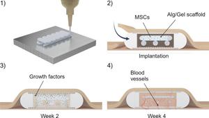
Subcutaneous delivery of cell therapy is an appealing minimally-invasive strategy for the treatment of various diseases. However, the subdermal site is poorly vascularized making it inadequate for supporting engraftment, viability, and function of exogenous cells. In this study, we developed a 3D bioprinted scaffold composed of alginate/gelatin (Alg/Gel) embedded with mesenchymal stem cells (MSCs) to enhance vascularization and tissue ingrowth in a subcutaneous microenvironment. We identified bio-ink crosslinking conditions that optimally recapitulated the mechanical properties of subcutaneous tissue. We achieved controlled degradation of the Alg/Gel scaffold synchronous with host tissue ingrowth and remodeling. Further, in a rat model, the Alg/Gel scaffold was superior to MSC-embedded Pluronic hydrogel in supporting tissue development and vascularization of a subcutaneous site. While the scaffold alone promoted vascular tissue formation, the inclusion of MSCs in the bio-ink further enhanced angiogenesis. Our findings highlight the use of simple cell-laden degradable bioprinted structures to generate a supportive microenvironment for cell delivery.
期刊介绍:
Biomedical Microdevices: BioMEMS and Biomedical Nanotechnology is an interdisciplinary periodical devoted to all aspects of research in the medical diagnostic and therapeutic applications of Micro-Electro-Mechanical Systems (BioMEMS) and nanotechnology for medicine and biology.
General subjects of interest include the design, characterization, testing, modeling and clinical validation of microfabricated systems, and their integration on-chip and in larger functional units. The specific interests of the Journal include systems for neural stimulation and recording, bioseparation technologies such as nanofilters and electrophoretic equipment, miniaturized analytic and DNA identification systems, biosensors, and micro/nanotechnologies for cell and tissue research, tissue engineering, cell transplantation, and the controlled release of drugs and biological molecules.
Contributions reporting on fundamental and applied investigations of the material science, biochemistry, and physics of biomedical microdevices and nanotechnology are encouraged. A non-exhaustive list of fields of interest includes: nanoparticle synthesis, characterization, and validation of therapeutic or imaging efficacy in animal models; biocompatibility; biochemical modification of microfabricated devices, with reference to non-specific protein adsorption, and the active immobilization and patterning of proteins on micro/nanofabricated surfaces; the dynamics of fluids in micro-and-nano-fabricated channels; the electromechanical and structural response of micro/nanofabricated systems; the interactions of microdevices with cells and tissues, including biocompatibility and biodegradation studies; variations in the characteristics of the systems as a function of the micro/nanofabrication parameters.


 分享
分享
 求助内容:
求助内容: 应助结果提醒方式:
 扫码关注我们
