Natasha Vassileff, Jereme G. Spiers, Sarah E. Bamford, Rohan G. T. Lowe, Keshava K. Datta, Paul J. Pigram, Andrew F. Hill
{"title":"Microglial activation induces nitric oxide signalling and alters protein S-nitrosylation patterns in extracellular vesicles","authors":"Natasha Vassileff, Jereme G. Spiers, Sarah E. Bamford, Rohan G. T. Lowe, Keshava K. Datta, Paul J. Pigram, Andrew F. Hill","doi":"10.1002/jev2.12455","DOIUrl":null,"url":null,"abstract":"<p>Neuroinflammation is an underlying feature of neurodegenerative conditions, often appearing early in the aetiology of a disease. Microglial activation, a prominent initiator of neuroinflammation, can be induced through lipopolysaccharide (LPS) treatment resulting in expression of the inducible form of nitric oxide synthase (iNOS), which produces nitric oxide (NO). NO post-translationally modifies cysteine thiols through S-nitrosylation, which can alter function of the target protein. Furthermore, packaging of these NO-modified proteins into extracellular vesicles (EVs) allows for the exertion of NO signalling in distant locations, resulting in further propagation of the neuroinflammatory phenotype. Despite this, the NO-modified proteome of activated microglial EVs has not been investigated. This study aimed to identify the protein post-translational modifications NO signalling induces in neuroinflammation. EVs isolated from LPS-treated microglia underwent mass spectral surface imaging using time of flight-secondary ion mass spectrometry (ToF-SIMS), in addition to iodolabelling and comparative proteomic analysis to identify post-translation S-nitrosylation modifications. ToF-SIMS imaging successfully identified cysteine thiol side chains modified through NO signalling in the LPS treated microglial-derived EV proteins. In addition, the iodolabelling proteomic analysis revealed that the EVs from LPS-treated microglia carried S-nitrosylated proteins indicative of neuroinflammation. These included known NO-modified proteins and those associated with LPS-induced microglial activation that may play an essential role in neuroinflammatory communication. Together, these results show activated microglia can exert broad NO signalling changes through the selective packaging of EVs during neuroinflammation.</p>","PeriodicalId":15811,"journal":{"name":"Journal of Extracellular Vesicles","volume":"13 6","pages":""},"PeriodicalIF":14.5000,"publicationDate":"2024-06-17","publicationTypes":"Journal Article","fieldsOfStudy":null,"isOpenAccess":false,"openAccessPdf":"https://onlinelibrary.wiley.com/doi/epdf/10.1002/jev2.12455","citationCount":"0","resultStr":null,"platform":"Semanticscholar","paperid":null,"PeriodicalName":"Journal of Extracellular Vesicles","FirstCategoryId":"3","ListUrlMain":"https://isevjournals.onlinelibrary.wiley.com/doi/10.1002/jev2.12455","RegionNum":1,"RegionCategory":"医学","ArticlePicture":[],"TitleCN":null,"AbstractTextCN":null,"PMCID":null,"EPubDate":"","PubModel":"","JCR":"Q1","JCRName":"CELL BIOLOGY","Score":null,"Total":0}
引用次数: 0
Abstract
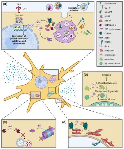
Neuroinflammation is an underlying feature of neurodegenerative conditions, often appearing early in the aetiology of a disease. Microglial activation, a prominent initiator of neuroinflammation, can be induced through lipopolysaccharide (LPS) treatment resulting in expression of the inducible form of nitric oxide synthase (iNOS), which produces nitric oxide (NO). NO post-translationally modifies cysteine thiols through S-nitrosylation, which can alter function of the target protein. Furthermore, packaging of these NO-modified proteins into extracellular vesicles (EVs) allows for the exertion of NO signalling in distant locations, resulting in further propagation of the neuroinflammatory phenotype. Despite this, the NO-modified proteome of activated microglial EVs has not been investigated. This study aimed to identify the protein post-translational modifications NO signalling induces in neuroinflammation. EVs isolated from LPS-treated microglia underwent mass spectral surface imaging using time of flight-secondary ion mass spectrometry (ToF-SIMS), in addition to iodolabelling and comparative proteomic analysis to identify post-translation S-nitrosylation modifications. ToF-SIMS imaging successfully identified cysteine thiol side chains modified through NO signalling in the LPS treated microglial-derived EV proteins. In addition, the iodolabelling proteomic analysis revealed that the EVs from LPS-treated microglia carried S-nitrosylated proteins indicative of neuroinflammation. These included known NO-modified proteins and those associated with LPS-induced microglial activation that may play an essential role in neuroinflammatory communication. Together, these results show activated microglia can exert broad NO signalling changes through the selective packaging of EVs during neuroinflammation.
期刊介绍:
The Journal of Extracellular Vesicles is an open access research publication that focuses on extracellular vesicles, including microvesicles, exosomes, ectosomes, and apoptotic bodies. It serves as the official journal of the International Society for Extracellular Vesicles and aims to facilitate the exchange of data, ideas, and information pertaining to the chemistry, biology, and applications of extracellular vesicles. The journal covers various aspects such as the cellular and molecular mechanisms of extracellular vesicles biogenesis, technological advancements in their isolation, quantification, and characterization, the role and function of extracellular vesicles in biology, stem cell-derived extracellular vesicles and their biology, as well as the application of extracellular vesicles for pharmacological, immunological, or genetic therapies.
The Journal of Extracellular Vesicles is widely recognized and indexed by numerous services, including Biological Abstracts, BIOSIS Previews, Chemical Abstracts Service (CAS), Current Contents/Life Sciences, Directory of Open Access Journals (DOAJ), Journal Citation Reports/Science Edition, Google Scholar, ProQuest Natural Science Collection, ProQuest SciTech Collection, SciTech Premium Collection, PubMed Central/PubMed, Science Citation Index Expanded, ScienceOpen, and Scopus.


 分享
分享
 求助内容:
求助内容: 应助结果提醒方式:
 扫码关注我们
