{"title":"An unusual case of nodular fasciitis presenting as an intra-tendinous mass.","authors":"Sisith Ariyaratne, Adesegun Abudu, Vaiyapuri Sumathi, Rajesh Botchu, Christine Azzopardi","doi":"10.1007/s00256-024-04728-x","DOIUrl":null,"url":null,"abstract":"<p><p>Nodular fasciitis is a benign soft tissue pseudotumour typically occurring in the upper extremities, head and neck, thigh and trunk. It is most commonly seen in subcutaneous locations but also can be present in intramuscular and intermuscular (fascial) locations. Its occurrence in the hand is rare, and while it can occur in close proximity to tendons, its presentation as an intra-tendinous mass has not been previously described. We present a unique and rare case of nodular fasciitis arising within the flexor digitorum profundus (FDP) tendon of the hand in a 16-year-old female. The patient presented with a painful swelling in the volar aspect of the base of her left middle finger, with progressive flexion deformity of the finger. Ultrasound and magnetic resonance imaging revealed a mass within the FDP tendon of the middle finger. An ultrasound-guided biopsy revealed a diagnosis of nodular fasciitis. Given the self-limiting nature of the condition, she was managed conservatively with close clinical and imaging follow-up. This case highlights the importance of considering nodular fasciitis in the differential diagnosis of an intra-tendinous lesion in the hand, even though it is a rare occurrence in this location. The clinical presentation, diagnostic workup, and management of this unique case are discussed, emphasising the potential for its misdiagnosis as a malignancy which can have important implications in management.</p>","PeriodicalId":21783,"journal":{"name":"Skeletal Radiology","volume":" ","pages":"367-371"},"PeriodicalIF":1.9000,"publicationDate":"2025-02-01","publicationTypes":"Journal Article","fieldsOfStudy":null,"isOpenAccess":false,"openAccessPdf":"","citationCount":"0","resultStr":null,"platform":"Semanticscholar","paperid":null,"PeriodicalName":"Skeletal Radiology","FirstCategoryId":"3","ListUrlMain":"https://doi.org/10.1007/s00256-024-04728-x","RegionNum":3,"RegionCategory":"医学","ArticlePicture":[],"TitleCN":null,"AbstractTextCN":null,"PMCID":null,"EPubDate":"2024/6/18 0:00:00","PubModel":"Epub","JCR":"Q2","JCRName":"ORTHOPEDICS","Score":null,"Total":0}
引用次数: 0
Abstract
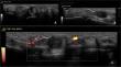
Nodular fasciitis is a benign soft tissue pseudotumour typically occurring in the upper extremities, head and neck, thigh and trunk. It is most commonly seen in subcutaneous locations but also can be present in intramuscular and intermuscular (fascial) locations. Its occurrence in the hand is rare, and while it can occur in close proximity to tendons, its presentation as an intra-tendinous mass has not been previously described. We present a unique and rare case of nodular fasciitis arising within the flexor digitorum profundus (FDP) tendon of the hand in a 16-year-old female. The patient presented with a painful swelling in the volar aspect of the base of her left middle finger, with progressive flexion deformity of the finger. Ultrasound and magnetic resonance imaging revealed a mass within the FDP tendon of the middle finger. An ultrasound-guided biopsy revealed a diagnosis of nodular fasciitis. Given the self-limiting nature of the condition, she was managed conservatively with close clinical and imaging follow-up. This case highlights the importance of considering nodular fasciitis in the differential diagnosis of an intra-tendinous lesion in the hand, even though it is a rare occurrence in this location. The clinical presentation, diagnostic workup, and management of this unique case are discussed, emphasising the potential for its misdiagnosis as a malignancy which can have important implications in management.
期刊介绍:
Skeletal Radiology provides a forum for the dissemination of current knowledge and information dealing with disorders of the musculoskeletal system including the spine. While emphasizing the radiological aspects of the many varied skeletal abnormalities, the journal also adopts an interdisciplinary approach, reflecting the membership of the International Skeletal Society. Thus, the anatomical, pathological, physiological, clinical, metabolic and epidemiological aspects of the many entities affecting the skeleton receive appropriate consideration.
This is the Journal of the International Skeletal Society and the Official Journal of the Society of Skeletal Radiology and the Australasian Musculoskelelal Imaging Group.


 分享
分享
 求助内容:
求助内容: 应助结果提醒方式:
 扫码关注我们
