{"title":"Imaging kidney inflammation using an oxidatively activated MRI probe","authors":"","doi":"10.1016/j.kint.2024.05.027","DOIUrl":null,"url":null,"abstract":"<div><p><span>Imaging tools for kidney inflammation<span><span> could improve care for patients suffering inflammatory kidney diseases by lessening reliance on </span>percutaneous biopsy<span> or biochemical tests alone. During kidney inflammation, infiltration of myeloid </span></span></span>immune cells<span><span><span><span> generates a kidney microenvironment that is oxidizing relative to normal kidney. Here, we evaluated whether magnetic resonance imaging (MRI) using the redox-active iron (Fe) complex Fe-PyC3A as an oxidatively activated probe could serve as a marker of kidney inflammation using mouse models of unilateral ischemia-reperfusion injury (IRI) and lupus nephritis<span> (MRL-lpr mice). We imaged unilateral IRI in gp91phox knockout mice, which are deficient in the </span></span>nicotinamide </span>oxidase<span> II (NOX2) enzyme required for myeloid oxidative burst, as loss of function control, and imaged MRL/MpJ mice as non-kidney involved lupus control. Gadoterate meglumine was used as a non-oxidatively activated control MRI probe. Fe-PyC3A safety was preliminarily examined following a single acute dose. Fe-PyC3A generated significantly greater MRI signal enhancement in the IRI kidney compared to the </span></span>contralateral<span> kidney in wild-type mice, but the effect was not observed in the NOX2-deficient control. Fe-PyC3A also generated significantly greater kidney enhancement in MRL-lpr mice compared to MRL/MpJ control. Gadoterate meglumine did not differentially enhance the IRI kidney over the contralateral kidney and did not differentially enhance the kidneys of MRL-lpr over MRL/MpJ mice. Fe-PyC3A was well tolerated at the highest dose evaluated, which was a 40-fold greater than required for imaging. Thus, our data indicate that MRI using Fe-PyC3A is specific to an oxidizing kidney environment shaped by activity of myeloid immune cells and support further evaluation of Fe-PyC3A for imaging kidney inflammation.</span></span></p></div>","PeriodicalId":17801,"journal":{"name":"Kidney international","volume":"106 4","pages":"Pages 671-678"},"PeriodicalIF":12.6000,"publicationDate":"2024-10-01","publicationTypes":"Journal Article","fieldsOfStudy":null,"isOpenAccess":false,"openAccessPdf":"","citationCount":"0","resultStr":null,"platform":"Semanticscholar","paperid":null,"PeriodicalName":"Kidney international","FirstCategoryId":"3","ListUrlMain":"https://www.sciencedirect.com/science/article/pii/S0085253824004083","RegionNum":1,"RegionCategory":"医学","ArticlePicture":[],"TitleCN":null,"AbstractTextCN":null,"PMCID":null,"EPubDate":"2024/6/18 0:00:00","PubModel":"Epub","JCR":"Q1","JCRName":"UROLOGY & NEPHROLOGY","Score":null,"Total":0}
引用次数: 0
Abstract
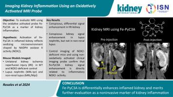
Imaging tools for kidney inflammation could improve care for patients suffering inflammatory kidney diseases by lessening reliance on percutaneous biopsy or biochemical tests alone. During kidney inflammation, infiltration of myeloid immune cells generates a kidney microenvironment that is oxidizing relative to normal kidney. Here, we evaluated whether magnetic resonance imaging (MRI) using the redox-active iron (Fe) complex Fe-PyC3A as an oxidatively activated probe could serve as a marker of kidney inflammation using mouse models of unilateral ischemia-reperfusion injury (IRI) and lupus nephritis (MRL-lpr mice). We imaged unilateral IRI in gp91phox knockout mice, which are deficient in the nicotinamide oxidase II (NOX2) enzyme required for myeloid oxidative burst, as loss of function control, and imaged MRL/MpJ mice as non-kidney involved lupus control. Gadoterate meglumine was used as a non-oxidatively activated control MRI probe. Fe-PyC3A safety was preliminarily examined following a single acute dose. Fe-PyC3A generated significantly greater MRI signal enhancement in the IRI kidney compared to the contralateral kidney in wild-type mice, but the effect was not observed in the NOX2-deficient control. Fe-PyC3A also generated significantly greater kidney enhancement in MRL-lpr mice compared to MRL/MpJ control. Gadoterate meglumine did not differentially enhance the IRI kidney over the contralateral kidney and did not differentially enhance the kidneys of MRL-lpr over MRL/MpJ mice. Fe-PyC3A was well tolerated at the highest dose evaluated, which was a 40-fold greater than required for imaging. Thus, our data indicate that MRI using Fe-PyC3A is specific to an oxidizing kidney environment shaped by activity of myeloid immune cells and support further evaluation of Fe-PyC3A for imaging kidney inflammation.
期刊介绍:
Kidney International (KI), the official journal of the International Society of Nephrology, is led by Dr. Pierre Ronco (Paris, France) and stands as one of nephrology's most cited and esteemed publications worldwide.
KI provides exceptional benefits for both readers and authors, featuring highly cited original articles, focused reviews, cutting-edge imaging techniques, and lively discussions on controversial topics.
The journal is dedicated to kidney research, serving researchers, clinical investigators, and practicing nephrologists.


 分享
分享
 求助内容:
求助内容: 应助结果提醒方式:
 扫码关注我们
