Woheeb M Saeed, Jude K Yoshino, Alexandria J Traynham, Nathaniel M Fried
{"title":"Simultaneous sealing and bisection of porcine renal blood vessels, ex vivo, using a continuous-wave, infrared diode laser at 1470 nm.","authors":"Woheeb M Saeed, Jude K Yoshino, Alexandria J Traynham, Nathaniel M Fried","doi":"10.1007/s10103-024-04093-0","DOIUrl":null,"url":null,"abstract":"<p><p>Electrosurgical and ultrasonic devices are used in surgical procedures for hemostatic sealing and bisection of vascular tissues. Previous benchtop studies alternatively demonstrated successful infrared laser sealing and cutting of blood vessels, in a sequential, two-step approach. This study describes a smaller, laparoscopic device compatible design, and simultaneous approach to sealing and bisection of vessels, with potential optical feedback. A 1470-nm infrared diode laser sealed and bisected 40 porcine renal arteries, ex vivo. A reciprocating, side-firing, optical fiber, housed in a transparent square quartz optical chamber (2.7 × 2.7 × 25 mm outer dimensions), delivered laser energy over an 11 mm scan length, with a range of incident powers (41-59 W) and treatment times (5-21 s). Vessel diameters ranged from 2.5 to 4.8 mm. Vessel burst pressure measurements were performed on each cut end (n = 80) with success indicated by pressures exceeding 360 mmHg. All vessel ends were successfully sealed and bisected (80/80). The highest incident power, 59 W, yielded short treatment times of 5-6 s. Peak temperatures on the external chamber surface reached 103 <sup>o</sup>C. Time to cool down to body temperature measured 37 s. Infrared lasers simultaneously seal and bisect blood vessels, with treatment times comparable to, and temperatures and cooling times lower than reported for conventional devices. Future work will focus on integrating the fiber and chamber into a standard 5-mm-outer-diameter laparoscopic device. Customization of fiber scan length to match vessel size may also reduce laser energy deposition, enabling lower peak temperatures, treatment times, and cooling times.</p>","PeriodicalId":17978,"journal":{"name":"Lasers in Medical Science","volume":"39 1","pages":"161"},"PeriodicalIF":2.4000,"publicationDate":"2024-06-22","publicationTypes":"Journal Article","fieldsOfStudy":null,"isOpenAccess":false,"openAccessPdf":"https://www.ncbi.nlm.nih.gov/pmc/articles/PMC11192808/pdf/","citationCount":"0","resultStr":null,"platform":"Semanticscholar","paperid":null,"PeriodicalName":"Lasers in Medical Science","FirstCategoryId":"5","ListUrlMain":"https://doi.org/10.1007/s10103-024-04093-0","RegionNum":4,"RegionCategory":"医学","ArticlePicture":[],"TitleCN":null,"AbstractTextCN":null,"PMCID":null,"EPubDate":"","PubModel":"","JCR":"Q3","JCRName":"ENGINEERING, BIOMEDICAL","Score":null,"Total":0}
引用次数: 0
Abstract
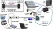
Electrosurgical and ultrasonic devices are used in surgical procedures for hemostatic sealing and bisection of vascular tissues. Previous benchtop studies alternatively demonstrated successful infrared laser sealing and cutting of blood vessels, in a sequential, two-step approach. This study describes a smaller, laparoscopic device compatible design, and simultaneous approach to sealing and bisection of vessels, with potential optical feedback. A 1470-nm infrared diode laser sealed and bisected 40 porcine renal arteries, ex vivo. A reciprocating, side-firing, optical fiber, housed in a transparent square quartz optical chamber (2.7 × 2.7 × 25 mm outer dimensions), delivered laser energy over an 11 mm scan length, with a range of incident powers (41-59 W) and treatment times (5-21 s). Vessel diameters ranged from 2.5 to 4.8 mm. Vessel burst pressure measurements were performed on each cut end (n = 80) with success indicated by pressures exceeding 360 mmHg. All vessel ends were successfully sealed and bisected (80/80). The highest incident power, 59 W, yielded short treatment times of 5-6 s. Peak temperatures on the external chamber surface reached 103 oC. Time to cool down to body temperature measured 37 s. Infrared lasers simultaneously seal and bisect blood vessels, with treatment times comparable to, and temperatures and cooling times lower than reported for conventional devices. Future work will focus on integrating the fiber and chamber into a standard 5-mm-outer-diameter laparoscopic device. Customization of fiber scan length to match vessel size may also reduce laser energy deposition, enabling lower peak temperatures, treatment times, and cooling times.
期刊介绍:
Lasers in Medical Science (LIMS) has established itself as the leading international journal in the rapidly expanding field of medical and dental applications of lasers and light. It provides a forum for the publication of papers on the technical, experimental, and clinical aspects of the use of medical lasers, including lasers in surgery, endoscopy, angioplasty, hyperthermia of tumors, and photodynamic therapy. In addition to medical laser applications, LIMS presents high-quality manuscripts on a wide range of dental topics, including aesthetic dentistry, endodontics, orthodontics, and prosthodontics.
The journal publishes articles on the medical and dental applications of novel laser technologies, light delivery systems, sensors to monitor laser effects, basic laser-tissue interactions, and the modeling of laser-tissue interactions. Beyond laser applications, LIMS features articles relating to the use of non-laser light-tissue interactions.


 分享
分享
 求助内容:
求助内容: 应助结果提醒方式:
 扫码关注我们
