Aysun Patterson, Mehmet Cengiz Karaismailoğlu, Orhan Küçüker
{"title":"Seed morphology of 31 Euphorbia L. species (Euphorbiaceae) in Turkey and their taxonomic significance","authors":"Aysun Patterson, Mehmet Cengiz Karaismailoğlu, Orhan Küçüker","doi":"10.1002/jemt.24637","DOIUrl":null,"url":null,"abstract":"<div>\n \n \n <section>\n \n <p>This paper includes a comprehensive taxonomical study based on seed morphology of 31 <i>Euphorbia L</i> species from Türkiye. The studied <i>Euphorbia</i> taxa have been examined for morphological traits such as seed color, dimensions, surface ornamentation, cell wall structures, lipid granule presence, and caruncle shape and dimensions with Scanning electron microscopy (SEM) and stereo microscopy to develop a better understanding of the basis of its species. The outcomes show that the species differ based on seed shape and color. The seed width dimensions are between 0.55 and 3.83 mm and the length dimensions are between 1.03 and 5.87 mm. <i>Euphorbia lathyris</i>, <i>E. prostrata</i>, and <i>E. nutans</i> are marked differently from the rest of the studied species based on their seed dimension. The seed surface ornamentation is classified into 12 different types: tuberculate, reticulate, areolate, colliculate, verrucate, alveolate, rugose, alveolate-reticulate, slightly reticulate, reticulate-areolate, pusticulate, and ruminate. The most common form is reticulate, found in eight species. The tuberculate (in <i>E. helioscopia</i>), areolate (in <i>E. oblongata</i>), slightly reticulate (in <i>E. amygdaloides</i>), and ruminate (in <i>E. herniariifolia</i>) ornamentation types are each characterized by only one species. The presence of lipid granules and anticlinal and periclinal cell walls disclose interspecific relationships within the examined taxa. Also, an identification key is offered for the studied species based on seed characters.</p>\n </section>\n \n <section>\n \n <h3> Research Highlights</h3>\n \n <div>\n <ul>\n \n <li>The seeds of Turkish <i>Euphorbia</i> species have been studied in depth.</li>\n \n <li>The morphological characters of seeds of Turkish <i>Euphorbia</i> species have been examined utilizing SEM and light microscopy for the first time and discussed the taxonomic practice of these characteristics.</li>\n \n <li>A dichotomous key containing seed morphological data has presented.</li>\n </ul>\n </div>\n </section>\n </div>","PeriodicalId":18684,"journal":{"name":"Microscopy Research and Technique","volume":"87 11","pages":"2654-2665"},"PeriodicalIF":2.1000,"publicationDate":"2024-06-25","publicationTypes":"Journal Article","fieldsOfStudy":null,"isOpenAccess":false,"openAccessPdf":"","citationCount":"0","resultStr":null,"platform":"Semanticscholar","paperid":null,"PeriodicalName":"Microscopy Research and Technique","FirstCategoryId":"5","ListUrlMain":"https://analyticalsciencejournals.onlinelibrary.wiley.com/doi/10.1002/jemt.24637","RegionNum":3,"RegionCategory":"工程技术","ArticlePicture":[],"TitleCN":null,"AbstractTextCN":null,"PMCID":null,"EPubDate":"","PubModel":"","JCR":"Q2","JCRName":"ANATOMY & MORPHOLOGY","Score":null,"Total":0}
引用次数: 0
Abstract
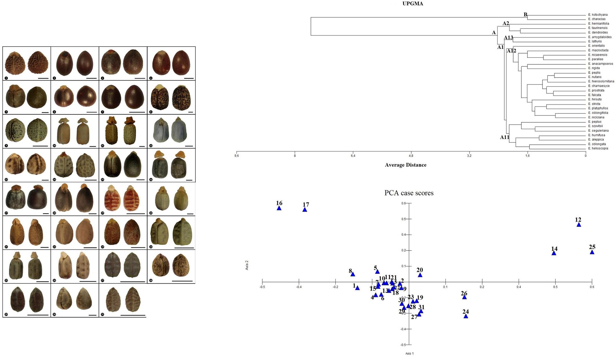
This paper includes a comprehensive taxonomical study based on seed morphology of 31 Euphorbia L species from Türkiye. The studied Euphorbia taxa have been examined for morphological traits such as seed color, dimensions, surface ornamentation, cell wall structures, lipid granule presence, and caruncle shape and dimensions with Scanning electron microscopy (SEM) and stereo microscopy to develop a better understanding of the basis of its species. The outcomes show that the species differ based on seed shape and color. The seed width dimensions are between 0.55 and 3.83 mm and the length dimensions are between 1.03 and 5.87 mm. Euphorbia lathyris, E. prostrata, and E. nutans are marked differently from the rest of the studied species based on their seed dimension. The seed surface ornamentation is classified into 12 different types: tuberculate, reticulate, areolate, colliculate, verrucate, alveolate, rugose, alveolate-reticulate, slightly reticulate, reticulate-areolate, pusticulate, and ruminate. The most common form is reticulate, found in eight species. The tuberculate (in E. helioscopia), areolate (in E. oblongata), slightly reticulate (in E. amygdaloides), and ruminate (in E. herniariifolia) ornamentation types are each characterized by only one species. The presence of lipid granules and anticlinal and periclinal cell walls disclose interspecific relationships within the examined taxa. Also, an identification key is offered for the studied species based on seed characters.
Research Highlights
The seeds of Turkish Euphorbia species have been studied in depth.
The morphological characters of seeds of Turkish Euphorbia species have been examined utilizing SEM and light microscopy for the first time and discussed the taxonomic practice of these characteristics.
A dichotomous key containing seed morphological data has presented.
期刊介绍:
Microscopy Research and Technique (MRT) publishes articles on all aspects of advanced microscopy original architecture and methodologies with applications in the biological, clinical, chemical, and materials sciences. Original basic and applied research as well as technical papers dealing with the various subsets of microscopy are encouraged. MRT is the right form for those developing new microscopy methods or using the microscope to answer key questions in basic and applied research.


 分享
分享
 求助内容:
求助内容: 应助结果提醒方式:
 扫码关注我们
