Carsen R McDaniel, Thomas M Johnson, Brian W Stancoven, Adam R Lincicum
{"title":"Distribution of the intraosseous branch of the posterior superior alveolar artery relative to the posterior maxillary teeth.","authors":"Carsen R McDaniel, Thomas M Johnson, Brian W Stancoven, Adam R Lincicum","doi":"10.5624/isd.20230160","DOIUrl":null,"url":null,"abstract":"<p><strong>Purpose: </strong>Preoperative identification of the intraosseous posterior superior alveolar artery (PSAA) is critical when planning sinus surgery. This study was conducted to determine the distance between the cementoenamel junction and the PSAA, as well as to identify factors influencing the detection of the PSAA on cone-beam computed tomography (CBCT).</p><p><strong>Materials and methods: </strong>In total, 254 CBCT scans of maxillary sinuses, acquired with 2 different scanners, were examined to identify the PSAA. The distance from the cementoenamel junction (CEJ) to the PSAA was recorded at each maxillary posterior tooth position. Binomial logistic regression and multiple linear regression were employed to evaluate the effects of scanner type, CBCT parameters, sex, and age on PSAA detection and CEJ-PSAA distance, respectively. <i>P</i>-values less than 0.05 were considered to indicate statistical significance.</p><p><strong>Results: </strong>The mean CEJ-PSAA distances at the second molar, first molar, second premolar, and first premolar positions were 17.0±4.0 mm, 21.8±4.1 mm, 19.5±4.7 mm, and 19.9±4.9 mm for scanner 1, respectively, and 17.3±3.5 mm, 16.9±4.3 mm, 18.5±4.1 mm, and 18.4±4.3 mm for scanner 2. No independent variable significantly influenced PSAA detection. However, tooth position (b=-0.67, <i>P</i><0.05) and scanner type (b=-1.3, <i>P</i><0.05) were significant predictors of CEJ-PSAA distance.</p><p><strong>Conclusion: </strong>CBCT-based estimates of CEJ-PSAA distance were comparable to those obtained in previous studies involving cadavers, CT, and CBCT. The type of CBCT scanner may slightly influence this measurement. No independent variable significantly impacted PSAA detection.</p>","PeriodicalId":51714,"journal":{"name":"Imaging Science in Dentistry","volume":"54 2","pages":"121-127"},"PeriodicalIF":2.1000,"publicationDate":"2024-06-01","publicationTypes":"Journal Article","fieldsOfStudy":null,"isOpenAccess":false,"openAccessPdf":"https://www.ncbi.nlm.nih.gov/pmc/articles/PMC11211025/pdf/","citationCount":"0","resultStr":null,"platform":"Semanticscholar","paperid":null,"PeriodicalName":"Imaging Science in Dentistry","FirstCategoryId":"1085","ListUrlMain":"https://doi.org/10.5624/isd.20230160","RegionNum":0,"RegionCategory":null,"ArticlePicture":[],"TitleCN":null,"AbstractTextCN":null,"PMCID":null,"EPubDate":"2024/4/2 0:00:00","PubModel":"Epub","JCR":"Q3","JCRName":"DENTISTRY, ORAL SURGERY & MEDICINE","Score":null,"Total":0}
引用次数: 0
Abstract
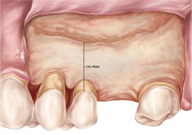
Purpose: Preoperative identification of the intraosseous posterior superior alveolar artery (PSAA) is critical when planning sinus surgery. This study was conducted to determine the distance between the cementoenamel junction and the PSAA, as well as to identify factors influencing the detection of the PSAA on cone-beam computed tomography (CBCT).
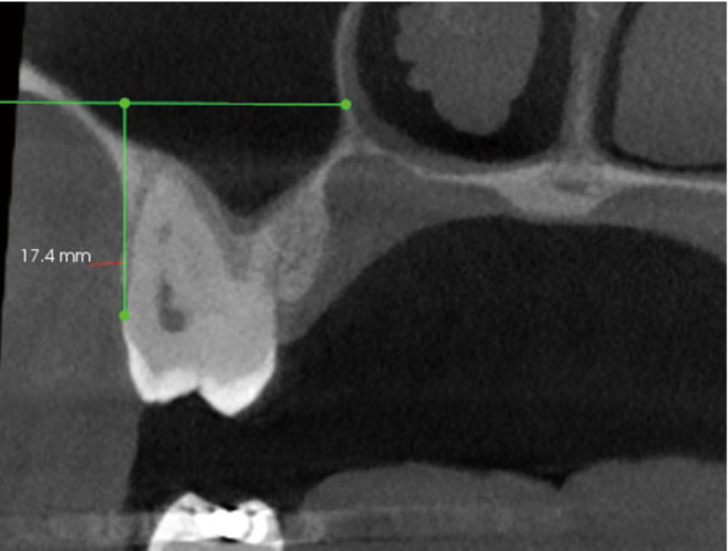
Materials and methods: In total, 254 CBCT scans of maxillary sinuses, acquired with 2 different scanners, were examined to identify the PSAA. The distance from the cementoenamel junction (CEJ) to the PSAA was recorded at each maxillary posterior tooth position. Binomial logistic regression and multiple linear regression were employed to evaluate the effects of scanner type, CBCT parameters, sex, and age on PSAA detection and CEJ-PSAA distance, respectively. P-values less than 0.05 were considered to indicate statistical significance.
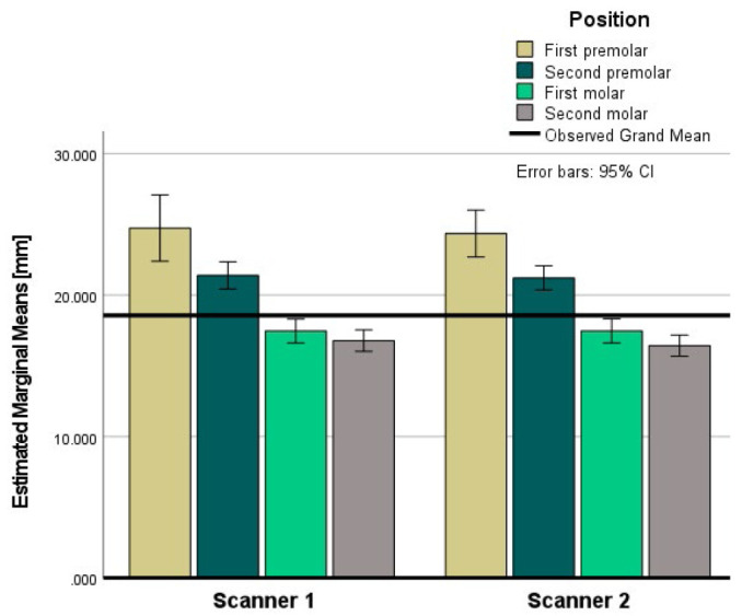
Results: The mean CEJ-PSAA distances at the second molar, first molar, second premolar, and first premolar positions were 17.0±4.0 mm, 21.8±4.1 mm, 19.5±4.7 mm, and 19.9±4.9 mm for scanner 1, respectively, and 17.3±3.5 mm, 16.9±4.3 mm, 18.5±4.1 mm, and 18.4±4.3 mm for scanner 2. No independent variable significantly influenced PSAA detection. However, tooth position (b=-0.67, P<0.05) and scanner type (b=-1.3, P<0.05) were significant predictors of CEJ-PSAA distance.
Conclusion: CBCT-based estimates of CEJ-PSAA distance were comparable to those obtained in previous studies involving cadavers, CT, and CBCT. The type of CBCT scanner may slightly influence this measurement. No independent variable significantly impacted PSAA detection.


 分享
分享
 求助内容:
求助内容: 应助结果提醒方式:
 扫码关注我们
