{"title":"Electrospun and 3D printed scaffolds based on biocompatible polymers for 3D cultivation of glioblastoma cells in vitro","authors":"R.A. Akasov , E.M. Trifanova , M.A. Khvorostina , A.V. Sochilina , S.A. Pavlova , A.I. Alekseeva , G.V. Pavlova , E.V. Khaydukov , V.K. Popov","doi":"10.1016/j.stlm.2024.100161","DOIUrl":null,"url":null,"abstract":"<div><p>Additive manufacturing techniques capable of fabricating biocompatible scaffolds with a given submicron/micron/supramicron structure are of growing interest for biomedical applications, including tissue engineering and tumor biology studies. Here, we propose antisolvent 3D printing and electrospinning techniques to obtain biopolymer scaffolds with different structural, mechanical, and surface properties to compare the cultivation patterns of glioblastoma cells. We found that human G01 cells, derived from human glioblastoma tumor tissue, were able to colonize the scaffolds in a time-dependent manner; the cells showed high viability as confirmed by colorimetric MTT assay, confocal fluorescence microscopy, and scanning electron microscopy data. Electrospun collagen scaffolds (low porosity, thin 2.75±0.22 μm fibers, low Young's modulus 0.076±0.033 MPa) provided monolayer-like growth of G01 glioblastoma cells with dense cell-cell contacts, while 3D-printed PLGA scaffolds (high porosity, thick ∼150 µm fibers, high Young's modulus 18±2 MPa) stimulated glioblastoma-specific spindle-like morphology. All scaffolds were non-toxic to cells and maintained cell growth for at least 2 weeks. The developed scaffolds could be further used for tumor research as a 3D model of glioblastoma <em>in vitro</em> or for tissue engineering of brain injury.</p></div>","PeriodicalId":72210,"journal":{"name":"Annals of 3D printed medicine","volume":"15 ","pages":"Article 100161"},"PeriodicalIF":0.0000,"publicationDate":"2024-08-01","publicationTypes":"Journal Article","fieldsOfStudy":null,"isOpenAccess":false,"openAccessPdf":"https://www.sciencedirect.com/science/article/pii/S2666964124000201/pdfft?md5=3296f0224a6061ad5aa5aced0dade772&pid=1-s2.0-S2666964124000201-main.pdf","citationCount":"0","resultStr":null,"platform":"Semanticscholar","paperid":null,"PeriodicalName":"Annals of 3D printed medicine","FirstCategoryId":"1085","ListUrlMain":"https://www.sciencedirect.com/science/article/pii/S2666964124000201","RegionNum":0,"RegionCategory":null,"ArticlePicture":[],"TitleCN":null,"AbstractTextCN":null,"PMCID":null,"EPubDate":"2024/6/22 0:00:00","PubModel":"Epub","JCR":"Q3","JCRName":"Medicine","Score":null,"Total":0}
引用次数: 0
Abstract
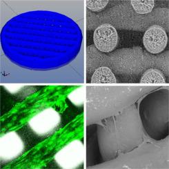
Additive manufacturing techniques capable of fabricating biocompatible scaffolds with a given submicron/micron/supramicron structure are of growing interest for biomedical applications, including tissue engineering and tumor biology studies. Here, we propose antisolvent 3D printing and electrospinning techniques to obtain biopolymer scaffolds with different structural, mechanical, and surface properties to compare the cultivation patterns of glioblastoma cells. We found that human G01 cells, derived from human glioblastoma tumor tissue, were able to colonize the scaffolds in a time-dependent manner; the cells showed high viability as confirmed by colorimetric MTT assay, confocal fluorescence microscopy, and scanning electron microscopy data. Electrospun collagen scaffolds (low porosity, thin 2.75±0.22 μm fibers, low Young's modulus 0.076±0.033 MPa) provided monolayer-like growth of G01 glioblastoma cells with dense cell-cell contacts, while 3D-printed PLGA scaffolds (high porosity, thick ∼150 µm fibers, high Young's modulus 18±2 MPa) stimulated glioblastoma-specific spindle-like morphology. All scaffolds were non-toxic to cells and maintained cell growth for at least 2 weeks. The developed scaffolds could be further used for tumor research as a 3D model of glioblastoma in vitro or for tissue engineering of brain injury.


 分享
分享
 求助内容:
求助内容: 应助结果提醒方式:
 扫码关注我们
