{"title":"Classification of variant portal vein anatomy based on three-dimensional CT: surgical implications.","authors":"Zheyu Liu, Tianni Shen, Kexin Xia, Junye He, Tianhao Rui, Wei Chen","doi":"10.1007/s00276-024-03427-5","DOIUrl":null,"url":null,"abstract":"<p><strong>Purposes: </strong>The purpose of this study was to develop a new and more comprehensive classification system for portal vein (PV) variations using three-dimensional visualization and evaluation (3DVE) and to discuss the prevalence rates and clinical implications of the variants.</p><p><strong>Methods: </strong>The anatomies of PVs were tracked and analyzed by using three-dimensional visualization of CT images acquired between 2013 and 2022. Scans from 200 adults were evaluated and a total of 178 patients (N = 178) were included in the study. The new classification system, named BLB classification, was developed based on the level of the absent PV branch in each variant anatomy.</p><p><strong>Results: </strong>Using the BLB classification system, PVs were divided into thirteen subtypes. Only 82.6-84.8% of the portal veins of the 178 patients were depicted in Atri's, Cheng's or Covey's classification, compared with 100% identified by the BLB classification. The BLB classification was validated against external data sets from previous studies, with 97.0-98.9% of patients classified by the BLB system.</p><p><strong>Conclusion: </strong>Variant PV anatomies are more commonly seen based on 3DVE than in previous reports. The BLB classification covers almost all portal vein variants and may be used for planning liver surgery.</p>","PeriodicalId":49461,"journal":{"name":"Surgical and Radiologic Anatomy","volume":null,"pages":null},"PeriodicalIF":1.4000,"publicationDate":"2024-08-01","publicationTypes":"Journal Article","fieldsOfStudy":null,"isOpenAccess":false,"openAccessPdf":"https://www.ncbi.nlm.nih.gov/pmc/articles/PMC11246292/pdf/","citationCount":"0","resultStr":null,"platform":"Semanticscholar","paperid":null,"PeriodicalName":"Surgical and Radiologic Anatomy","FirstCategoryId":"3","ListUrlMain":"https://doi.org/10.1007/s00276-024-03427-5","RegionNum":4,"RegionCategory":"医学","ArticlePicture":[],"TitleCN":null,"AbstractTextCN":null,"PMCID":null,"EPubDate":"2024/7/4 0:00:00","PubModel":"Epub","JCR":"Q2","JCRName":"Medicine","Score":null,"Total":0}
引用次数: 0
Abstract
Purposes: The purpose of this study was to develop a new and more comprehensive classification system for portal vein (PV) variations using three-dimensional visualization and evaluation (3DVE) and to discuss the prevalence rates and clinical implications of the variants.
Methods: The anatomies of PVs were tracked and analyzed by using three-dimensional visualization of CT images acquired between 2013 and 2022. Scans from 200 adults were evaluated and a total of 178 patients (N = 178) were included in the study. The new classification system, named BLB classification, was developed based on the level of the absent PV branch in each variant anatomy.
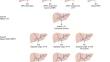
Results: Using the BLB classification system, PVs were divided into thirteen subtypes. Only 82.6-84.8% of the portal veins of the 178 patients were depicted in Atri's, Cheng's or Covey's classification, compared with 100% identified by the BLB classification. The BLB classification was validated against external data sets from previous studies, with 97.0-98.9% of patients classified by the BLB system.
Conclusion: Variant PV anatomies are more commonly seen based on 3DVE than in previous reports. The BLB classification covers almost all portal vein variants and may be used for planning liver surgery.
期刊介绍:
Anatomy is a morphological science which cannot fail to interest the clinician. The practical application of anatomical research to clinical problems necessitates special adaptation and selectivity in choosing from numerous international works. Although there is a tendency to believe that meaningful advances in anatomy are unlikely, constant revision is necessary. Surgical and Radiologic Anatomy, the first international journal of Clinical anatomy has been created in this spirit.
Its goal is to serve clinicians, regardless of speciality-physicians, surgeons, radiologists or other specialists-as an indispensable aid with which they can improve their knowledge of anatomy. Each issue includes: Original papers, review articles, articles on the anatomical bases of medical, surgical and radiological techniques, articles of normal radiologic anatomy, brief reviews of anatomical publications of clinical interest.
Particular attention is given to high quality illustrations, which are indispensable for a better understanding of anatomical problems.
Surgical and Radiologic Anatomy is a journal written by anatomists for clinicians with a special interest in anatomy.


 分享
分享
 求助内容:
求助内容: 应助结果提醒方式:
 扫码关注我们
