Katharina S. Höffgen , Jennifer Dabel , Christian P. Konken , Dominic A. Depke , Sven Hermann , Wolfgang Dörner , Sonja Schelhaas , Michael Schäfers , Henning D. Mootz
{"title":"Combining poly-epitope MoonTags and labeled nanobodies for signal amplification in cell-specific PET imaging in vivo","authors":"Katharina S. Höffgen , Jennifer Dabel , Christian P. Konken , Dominic A. Depke , Sven Hermann , Wolfgang Dörner , Sonja Schelhaas , Michael Schäfers , Henning D. Mootz","doi":"10.1016/j.nucmedbio.2024.108937","DOIUrl":null,"url":null,"abstract":"<div><p>Immunorecognition provides an excellent basis for targeted imaging techniques covering a wide range from basic research to diagnostics and from single cells to whole organisms. Fluorescence- or radioisotope-labeled antibodies, antibody fragments or nanobodies enable a direct signal readout upon binding and allow for versatile imaging from microscopy to whole-body imaging. However, as the signal intensity directly correlates with the number of labeled antibodies bound to their epitopes (1:1 binding), sensitivity for low-expressing epitopes can be limiting for visualization. For the first time, we developed poly-epitope tags with multiple copies (1 to 7) of a short peptide epitope, specifically the MoonTag, that are recognized by a labeled nanobody and aimed at signal amplification in microscopy and cell-specific PET imaging. In transiently transfected HeLa cells or stably transduced A4573 cells we characterized complex formation and <em>in vitro</em> signal amplification. Indeed, using fluorescently and radioactively labeled nanobodies we found an approximately linear signal amplification with increasing numbers of epitope copies <em>in vitro</em>. To test the poly-epitope approach <em>in vivo</em>, A4573 tumor cells were injected subcutaneously into the shoulder of NSG mice, with A4573 tumor cells expressing a poly-epitope of 7 MoonTags on one side and WT cells on the other side. Using a [<sup>68</sup>Ga]-labeled NODAGA-conjugated MoonTag nanobody, we performed PET/CT imaging at day 8–9 after tumor implantation. Specific binding of a [<sup>68</sup>Ga]-labeled NODAGA-conjugated MoonTag nanobody was observed in 7xMoonTag tumors (1.7 ± 0.5%ID/mL) by PET imaging, showing significantly higher radiotracer accumulation compared to the WT tumors (1.1 ± 0.3%ID/mL; <em>p</em> < 0.01). <em>Ex vivo</em> gamma counter measurements confirmed significantly higher uptake in 7xMoonTag tumors compared to WT tumors (<em>p</em> < 0.001). In addition, MoonTag nanobody binding was detected by autoradiography which was spatially matched with histological analysis of the tumor tissues. In conclusion, we expect nanobody-based poly-epitope tag strategies to be widely applicable for multimodal imaging techniques given the advantageous properties of nanobodies and their amenability to genetic and chemical engineering.</p></div>","PeriodicalId":19363,"journal":{"name":"Nuclear medicine and biology","volume":"136 ","pages":"Article 108937"},"PeriodicalIF":3.0000,"publicationDate":"2024-09-01","publicationTypes":"Journal Article","fieldsOfStudy":null,"isOpenAccess":false,"openAccessPdf":"https://www.sciencedirect.com/science/article/pii/S0969805124000635/pdfft?md5=538e2cf736ea2337611b25300b461524&pid=1-s2.0-S0969805124000635-main.pdf","citationCount":"0","resultStr":null,"platform":"Semanticscholar","paperid":null,"PeriodicalName":"Nuclear medicine and biology","FirstCategoryId":"3","ListUrlMain":"https://www.sciencedirect.com/science/article/pii/S0969805124000635","RegionNum":4,"RegionCategory":"医学","ArticlePicture":[],"TitleCN":null,"AbstractTextCN":null,"PMCID":null,"EPubDate":"2024/6/20 0:00:00","PubModel":"Epub","JCR":"Q1","JCRName":"RADIOLOGY, NUCLEAR MEDICINE & MEDICAL IMAGING","Score":null,"Total":0}
引用次数: 0
Abstract
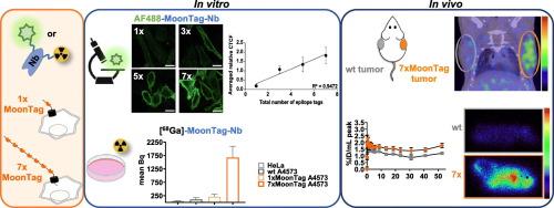
Immunorecognition provides an excellent basis for targeted imaging techniques covering a wide range from basic research to diagnostics and from single cells to whole organisms. Fluorescence- or radioisotope-labeled antibodies, antibody fragments or nanobodies enable a direct signal readout upon binding and allow for versatile imaging from microscopy to whole-body imaging. However, as the signal intensity directly correlates with the number of labeled antibodies bound to their epitopes (1:1 binding), sensitivity for low-expressing epitopes can be limiting for visualization. For the first time, we developed poly-epitope tags with multiple copies (1 to 7) of a short peptide epitope, specifically the MoonTag, that are recognized by a labeled nanobody and aimed at signal amplification in microscopy and cell-specific PET imaging. In transiently transfected HeLa cells or stably transduced A4573 cells we characterized complex formation and in vitro signal amplification. Indeed, using fluorescently and radioactively labeled nanobodies we found an approximately linear signal amplification with increasing numbers of epitope copies in vitro. To test the poly-epitope approach in vivo, A4573 tumor cells were injected subcutaneously into the shoulder of NSG mice, with A4573 tumor cells expressing a poly-epitope of 7 MoonTags on one side and WT cells on the other side. Using a [68Ga]-labeled NODAGA-conjugated MoonTag nanobody, we performed PET/CT imaging at day 8–9 after tumor implantation. Specific binding of a [68Ga]-labeled NODAGA-conjugated MoonTag nanobody was observed in 7xMoonTag tumors (1.7 ± 0.5%ID/mL) by PET imaging, showing significantly higher radiotracer accumulation compared to the WT tumors (1.1 ± 0.3%ID/mL; p < 0.01). Ex vivo gamma counter measurements confirmed significantly higher uptake in 7xMoonTag tumors compared to WT tumors (p < 0.001). In addition, MoonTag nanobody binding was detected by autoradiography which was spatially matched with histological analysis of the tumor tissues. In conclusion, we expect nanobody-based poly-epitope tag strategies to be widely applicable for multimodal imaging techniques given the advantageous properties of nanobodies and their amenability to genetic and chemical engineering.
期刊介绍:
Nuclear Medicine and Biology publishes original research addressing all aspects of radiopharmaceutical science: synthesis, in vitro and ex vivo studies, in vivo biodistribution by dissection or imaging, radiopharmacology, radiopharmacy, and translational clinical studies of new targeted radiotracers. The importance of the target to an unmet clinical need should be the first consideration. If the synthesis of a new radiopharmaceutical is submitted without in vitro or in vivo data, then the uniqueness of the chemistry must be emphasized.
These multidisciplinary studies should validate the mechanism of localization whether the probe is based on binding to a receptor, enzyme, tumor antigen, or another well-defined target. The studies should be aimed at evaluating how the chemical and radiopharmaceutical properties affect pharmacokinetics, pharmacodynamics, or therapeutic efficacy. Ideally, the study would address the sensitivity of the probe to changes in disease or treatment, although studies validating mechanism alone are acceptable. Radiopharmacy practice, addressing the issues of preparation, automation, quality control, dispensing, and regulations applicable to qualification and administration of radiopharmaceuticals to humans, is an important aspect of the developmental process, but only if the study has a significant impact on the field.
Contributions on the subject of therapeutic radiopharmaceuticals also are appropriate provided that the specificity of labeled compound localization and therapeutic effect have been addressed.


 分享
分享
 求助内容:
求助内容: 应助结果提醒方式:
 扫码关注我们
