Filippo Casoni, Laura Croci, Francesca Marroni, Giulia Demenego, Chiara Marullo, Ottavio Cremona, Franca Codazzi, G. Giacomo Consalez
{"title":"A spatial–temporal map of glutamatergic neurogenesis in the murine embryonic cerebellar nuclei uncovers a high degree of cellular heterogeneity","authors":"Filippo Casoni, Laura Croci, Francesca Marroni, Giulia Demenego, Chiara Marullo, Ottavio Cremona, Franca Codazzi, G. Giacomo Consalez","doi":"10.1111/joa.14107","DOIUrl":null,"url":null,"abstract":"<p>The nuclei are the main output structures of the cerebellum. Each and every cerebellar cortical computation reaches several areas of the brain by means of cerebellar nuclei processing and integration. Nevertheless, our knowledge of these structures is still limited compared to the cerebellar cortex. Here, we present a mouse genetic inducible fate-mapping study characterizing rhombic lip-derived glutamatergic neurons of the nuclei, the most conspicuous family of long-range cerebellar efferent neurons. Glutamatergic neurons mainly occupy dorsal and lateral territories of the lateral and interposed nuclei, as well as the entire medial nucleus. In mice, they are born starting from about embryonic day 9.5, with a peak between 10.5 and 12.5, and invade the nuclei with a lateral-to-medial progression. While some markers label a heterogeneous population of neurons sharing a common location (BRN2), others appear to be lineage specific (TBR1, LMX1a, and MEIS2). A comparative analysis of TBR1 and LMX1a distributions reveals an incomplete overlap in their expression domains, in keeping with the existence of separate efferent subpopulations. Finally, some tagged glutamatergic progenitors are not labeled by any of the markers used in this study, disclosing further complexity. Taken together, our results obtained in late embryonic nuclei shed light on the heterogeneity of the excitatory neuron pool, underlying the diversity in connectivity and functions of this largely unexplored cerebellar territory. Our findings contribute to laying the groundwork for a comprehensive functional analysis of nuclear neuron subpopulations.</p>","PeriodicalId":14971,"journal":{"name":"Journal of Anatomy","volume":"245 4","pages":"560-571"},"PeriodicalIF":1.9000,"publicationDate":"2024-07-06","publicationTypes":"Journal Article","fieldsOfStudy":null,"isOpenAccess":false,"openAccessPdf":"https://onlinelibrary.wiley.com/doi/epdf/10.1111/joa.14107","citationCount":"0","resultStr":null,"platform":"Semanticscholar","paperid":null,"PeriodicalName":"Journal of Anatomy","FirstCategoryId":"3","ListUrlMain":"https://onlinelibrary.wiley.com/doi/10.1111/joa.14107","RegionNum":3,"RegionCategory":"医学","ArticlePicture":[],"TitleCN":null,"AbstractTextCN":null,"PMCID":null,"EPubDate":"","PubModel":"","JCR":"Q2","JCRName":"ANATOMY & MORPHOLOGY","Score":null,"Total":0}
引用次数: 0
Abstract
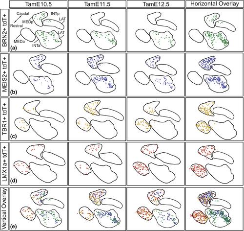
The nuclei are the main output structures of the cerebellum. Each and every cerebellar cortical computation reaches several areas of the brain by means of cerebellar nuclei processing and integration. Nevertheless, our knowledge of these structures is still limited compared to the cerebellar cortex. Here, we present a mouse genetic inducible fate-mapping study characterizing rhombic lip-derived glutamatergic neurons of the nuclei, the most conspicuous family of long-range cerebellar efferent neurons. Glutamatergic neurons mainly occupy dorsal and lateral territories of the lateral and interposed nuclei, as well as the entire medial nucleus. In mice, they are born starting from about embryonic day 9.5, with a peak between 10.5 and 12.5, and invade the nuclei with a lateral-to-medial progression. While some markers label a heterogeneous population of neurons sharing a common location (BRN2), others appear to be lineage specific (TBR1, LMX1a, and MEIS2). A comparative analysis of TBR1 and LMX1a distributions reveals an incomplete overlap in their expression domains, in keeping with the existence of separate efferent subpopulations. Finally, some tagged glutamatergic progenitors are not labeled by any of the markers used in this study, disclosing further complexity. Taken together, our results obtained in late embryonic nuclei shed light on the heterogeneity of the excitatory neuron pool, underlying the diversity in connectivity and functions of this largely unexplored cerebellar territory. Our findings contribute to laying the groundwork for a comprehensive functional analysis of nuclear neuron subpopulations.
期刊介绍:
Journal of Anatomy is an international peer-reviewed journal sponsored by the Anatomical Society. The journal publishes original papers, invited review articles and book reviews. Its main focus is to understand anatomy through an analysis of structure, function, development and evolution. Priority will be given to studies of that clearly articulate their relevance to the anatomical community. Focal areas include: experimental studies, contributions based on molecular and cell biology and on the application of modern imaging techniques and papers with novel methods or synthetic perspective on an anatomical system.
Studies that are essentially descriptive anatomy are appropriate only if they communicate clearly a broader functional or evolutionary significance. You must clearly state the broader implications of your work in the abstract.
We particularly welcome submissions in the following areas:
Cell biology and tissue architecture
Comparative functional morphology
Developmental biology
Evolutionary developmental biology
Evolutionary morphology
Functional human anatomy
Integrative vertebrate paleontology
Methodological innovations in anatomical research
Musculoskeletal system
Neuroanatomy and neurodegeneration
Significant advances in anatomical education.


 分享
分享
 求助内容:
求助内容: 应助结果提醒方式:
 扫码关注我们
