Paul Benjamin Klar, David Geoffrey Waterman, Tim Gruene, Debakshi Mullick, Yun Song, James Boris Gilchrist, C. David Owen, Wen Wen, Idan Biran, Lothar Houben, Neta Regev-Rudzki, Ron Dzikowski, Noa Marom, Lukas Palatinus, Peijun Zhang, Leslie Leiserowitz, Michael Elbaum
{"title":"Cryo-tomography and 3D Electron Diffraction Reveal the Polar Habit and Chiral Structure of the Malaria Pigment Crystal Hemozoin","authors":"Paul Benjamin Klar, David Geoffrey Waterman, Tim Gruene, Debakshi Mullick, Yun Song, James Boris Gilchrist, C. David Owen, Wen Wen, Idan Biran, Lothar Houben, Neta Regev-Rudzki, Ron Dzikowski, Noa Marom, Lukas Palatinus, Peijun Zhang, Leslie Leiserowitz, Michael Elbaum","doi":"10.1021/acscentsci.4c00162","DOIUrl":null,"url":null,"abstract":"Detoxification of heme in <i>Plasmodium</i> depends on its crystallization into hemozoin. This pathway is a major target of antimalarial drugs. The crystalline structure of hemozoin was established by X-ray powder diffraction using a synthetic analog, β-hematin. Here, we apply emerging methods of <i>in situ</i> cryo-electron tomography and 3D electron diffraction to obtain a definitive structure of hemozoin directly from ruptured parasite cells. Biogenic hemozoin crystals take a striking polar morphology. Like β-hematin, the unit cell contains a heme dimer, which may form four distinct stereoisomers: two centrosymmetric and two chiral enantiomers. Diffraction analysis, supported by density functional theory analysis, reveals a selective mixture in the hemozoin lattice of one centrosymmetric and one chiral dimer. Absolute configuration has been determined by morphological analysis and confirmed by a novel method of exit-wave reconstruction from a focal series. Atomic disorder appears on specific facets asymmetrically, and the polar morphology can be understood in light of water binding. Structural modeling of the heme detoxification protein suggests a function as a chiral agent to bias the dimer formation in favor of rapid growth of a single crystalline phase. The refined structure of hemozoin should serve as a guide to new drug development.","PeriodicalId":10,"journal":{"name":"ACS Central Science","volume":"34 1","pages":""},"PeriodicalIF":10.4000,"publicationDate":"2024-07-04","publicationTypes":"Journal Article","fieldsOfStudy":null,"isOpenAccess":false,"openAccessPdf":"","citationCount":"0","resultStr":null,"platform":"Semanticscholar","paperid":null,"PeriodicalName":"ACS Central Science","FirstCategoryId":"92","ListUrlMain":"https://doi.org/10.1021/acscentsci.4c00162","RegionNum":1,"RegionCategory":"化学","ArticlePicture":[],"TitleCN":null,"AbstractTextCN":null,"PMCID":null,"EPubDate":"","PubModel":"","JCR":"Q1","JCRName":"CHEMISTRY, MULTIDISCIPLINARY","Score":null,"Total":0}
引用次数: 0
Abstract
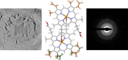
Detoxification of heme in Plasmodium depends on its crystallization into hemozoin. This pathway is a major target of antimalarial drugs. The crystalline structure of hemozoin was established by X-ray powder diffraction using a synthetic analog, β-hematin. Here, we apply emerging methods of in situ cryo-electron tomography and 3D electron diffraction to obtain a definitive structure of hemozoin directly from ruptured parasite cells. Biogenic hemozoin crystals take a striking polar morphology. Like β-hematin, the unit cell contains a heme dimer, which may form four distinct stereoisomers: two centrosymmetric and two chiral enantiomers. Diffraction analysis, supported by density functional theory analysis, reveals a selective mixture in the hemozoin lattice of one centrosymmetric and one chiral dimer. Absolute configuration has been determined by morphological analysis and confirmed by a novel method of exit-wave reconstruction from a focal series. Atomic disorder appears on specific facets asymmetrically, and the polar morphology can be understood in light of water binding. Structural modeling of the heme detoxification protein suggests a function as a chiral agent to bias the dimer formation in favor of rapid growth of a single crystalline phase. The refined structure of hemozoin should serve as a guide to new drug development.
疟原虫体内血红素的解毒依赖于其结晶成血色素。这一途径是抗疟药物的主要靶点。通过 X 射线粉末衍射,我们利用合成类似物 β-hematin,确定了安息香酸的晶体结构。在这里,我们应用新出现的原位低温电子断层扫描和三维电子衍射方法,直接从破裂的寄生虫细胞中获得了安息香血素的确定结构。生物造血素晶体具有显著的极性形态。与 β-血红素一样,其单胞包含一个血红素二聚体,可形成四种不同的立体异构体:两种中心对称异构体和两种手性对映体。在密度泛函理论分析的支持下,衍射分析揭示了血色素晶格中一种中心对称二聚体和一种手性二聚体的选择性混合物。绝对构型是通过形态分析确定的,并通过一种从焦点序列重建出口波的新方法得到了证实。原子紊乱不对称地出现在特定的面上,极性形态可以从水结合的角度来理解。血红素解毒蛋白的结构模型表明,它具有作为手性剂的功能,可偏向于二聚体的形成,从而有利于单晶相的快速生长。血色素的精制结构可作为新药开发的指南。
期刊介绍:
ACS Central Science publishes significant primary reports on research in chemistry and allied fields where chemical approaches are pivotal. As the first fully open-access journal by the American Chemical Society, it covers compelling and important contributions to the broad chemistry and scientific community. "Central science," a term popularized nearly 40 years ago, emphasizes chemistry's central role in connecting physical and life sciences, and fundamental sciences with applied disciplines like medicine and engineering. The journal focuses on exceptional quality articles, addressing advances in fundamental chemistry and interdisciplinary research.


 分享
分享
 求助内容:
求助内容: 应助结果提醒方式:
 扫码关注我们
