Sebastian M. Molnar, Yuriy Kim, Lindsay Wieczorek, Anastasia Williams, Kajal Ashok Patil, Pooja Khatkar, Mark F. Santos, Gifty Mensah, Aurelio Lorico, Victoria R. Polonis, Fatah Kashanchi
{"title":"Extracellular vesicle isolation methods identify distinct HIV-1 particles released from chronically infected T-cells","authors":"Sebastian M. Molnar, Yuriy Kim, Lindsay Wieczorek, Anastasia Williams, Kajal Ashok Patil, Pooja Khatkar, Mark F. Santos, Gifty Mensah, Aurelio Lorico, Victoria R. Polonis, Fatah Kashanchi","doi":"10.1002/jev2.12476","DOIUrl":null,"url":null,"abstract":"<p>The current study analyzed the intersecting biophysical, biochemical, and functional properties of extracellular particles (EPs) with the human immunodeficiency virus type-1 (HIV-1) beyond the currently accepted size range for HIV-1. We isolated five fractions (Frac-A through Frac-E) from HIV-infected cells by sequential differential ultracentrifugation (DUC). All fractions showed a heterogeneous size distribution with median particle sizes greater than 100 nm for Frac-A through Frac-D but not for Frac-E, which contained small EPs with an average size well below 50 nm. Synchronized and released cultures contained large infectious EPs in Frac-A, with markers of amphisomes and viral components. Additionally, Frac-E uniquely contained EPs positive for CD63, HSP70, and HIV-1 proteins. Despite its small average size, Frac-E contained membrane-protected viral integrase, detectable only after SDS treatment, indicating that it is enclosed in vesicles. Single particle analysis with dSTORM further supported these findings as CD63, HIV-1 integrase, and the viral surface envelope (Env) glycoprotein (gp) colocalized on the same Frac-E particles. Surprisingly, Frac-E EPs were infectious, and infectivity was significantly reduced by immunodepleting Frac-E with anti-CD63, indicating the presence of this protein on the surface of infectious small EPs in Frac-E. To our knowledge, this is the first time that extracellular vesicle (EV) isolation methods have identified infectious small HIV-1 particles (<sub>sm</sub>HIV-1) that are under 50 nm. Collectively, our data indicate that the crossroads between EPs and HIV-1 potentially extend beyond the currently accepted biophysical properties of HIV-1, which may have further implications for viral pathogenesis.</p>","PeriodicalId":15811,"journal":{"name":"Journal of Extracellular Vesicles","volume":"13 7","pages":""},"PeriodicalIF":14.5000,"publicationDate":"2024-07-08","publicationTypes":"Journal Article","fieldsOfStudy":null,"isOpenAccess":false,"openAccessPdf":"https://www.ncbi.nlm.nih.gov/pmc/articles/PMC11231049/pdf/","citationCount":"0","resultStr":null,"platform":"Semanticscholar","paperid":null,"PeriodicalName":"Journal of Extracellular Vesicles","FirstCategoryId":"3","ListUrlMain":"https://isevjournals.onlinelibrary.wiley.com/doi/10.1002/jev2.12476","RegionNum":1,"RegionCategory":"医学","ArticlePicture":[],"TitleCN":null,"AbstractTextCN":null,"PMCID":null,"EPubDate":"","PubModel":"","JCR":"Q1","JCRName":"CELL BIOLOGY","Score":null,"Total":0}
引用次数: 0
Abstract
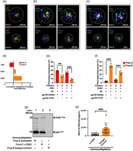
The current study analyzed the intersecting biophysical, biochemical, and functional properties of extracellular particles (EPs) with the human immunodeficiency virus type-1 (HIV-1) beyond the currently accepted size range for HIV-1. We isolated five fractions (Frac-A through Frac-E) from HIV-infected cells by sequential differential ultracentrifugation (DUC). All fractions showed a heterogeneous size distribution with median particle sizes greater than 100 nm for Frac-A through Frac-D but not for Frac-E, which contained small EPs with an average size well below 50 nm. Synchronized and released cultures contained large infectious EPs in Frac-A, with markers of amphisomes and viral components. Additionally, Frac-E uniquely contained EPs positive for CD63, HSP70, and HIV-1 proteins. Despite its small average size, Frac-E contained membrane-protected viral integrase, detectable only after SDS treatment, indicating that it is enclosed in vesicles. Single particle analysis with dSTORM further supported these findings as CD63, HIV-1 integrase, and the viral surface envelope (Env) glycoprotein (gp) colocalized on the same Frac-E particles. Surprisingly, Frac-E EPs were infectious, and infectivity was significantly reduced by immunodepleting Frac-E with anti-CD63, indicating the presence of this protein on the surface of infectious small EPs in Frac-E. To our knowledge, this is the first time that extracellular vesicle (EV) isolation methods have identified infectious small HIV-1 particles (smHIV-1) that are under 50 nm. Collectively, our data indicate that the crossroads between EPs and HIV-1 potentially extend beyond the currently accepted biophysical properties of HIV-1, which may have further implications for viral pathogenesis.
期刊介绍:
The Journal of Extracellular Vesicles is an open access research publication that focuses on extracellular vesicles, including microvesicles, exosomes, ectosomes, and apoptotic bodies. It serves as the official journal of the International Society for Extracellular Vesicles and aims to facilitate the exchange of data, ideas, and information pertaining to the chemistry, biology, and applications of extracellular vesicles. The journal covers various aspects such as the cellular and molecular mechanisms of extracellular vesicles biogenesis, technological advancements in their isolation, quantification, and characterization, the role and function of extracellular vesicles in biology, stem cell-derived extracellular vesicles and their biology, as well as the application of extracellular vesicles for pharmacological, immunological, or genetic therapies.
The Journal of Extracellular Vesicles is widely recognized and indexed by numerous services, including Biological Abstracts, BIOSIS Previews, Chemical Abstracts Service (CAS), Current Contents/Life Sciences, Directory of Open Access Journals (DOAJ), Journal Citation Reports/Science Edition, Google Scholar, ProQuest Natural Science Collection, ProQuest SciTech Collection, SciTech Premium Collection, PubMed Central/PubMed, Science Citation Index Expanded, ScienceOpen, and Scopus.


 分享
分享
 求助内容:
求助内容: 应助结果提醒方式:
 扫码关注我们
