Francesca Sbrana, Flaminia Chellini, Alessia Tani, Martina Parigi, Rachele Garella, Francesco Palmieri, Sandra Zecchi-Orlandini, Roberta Squecco, Chiara Sassoli
{"title":"Label-free three-dimensional imaging and quantitative analysis of living fibroblasts and myofibroblasts by holotomographic microscopy","authors":"Francesca Sbrana, Flaminia Chellini, Alessia Tani, Martina Parigi, Rachele Garella, Francesco Palmieri, Sandra Zecchi-Orlandini, Roberta Squecco, Chiara Sassoli","doi":"10.1002/jemt.24648","DOIUrl":null,"url":null,"abstract":"<div>\n \n \n <section>\n \n <p>Holotomography (HT) is a cutting-edge fast live-cell quantitative label-free imaging technique. Based on the principle of quantitative phase imaging, it combines holography and tomography to record a three-dimensional map of the refractive index, used as intrinsic optical and quantitative imaging contrast parameter of biological samples, at a sub-micrometer spatial resolution. In this study HT has been employed for the first time to analyze the changes of fibroblasts differentiating towards myofibroblasts – recognized as the main cell player of fibrosis – when cultured in vitro with the pro-fibrotic factor, namely transforming growth factor-β1. In parallel, F-actin, vinculin, α-smooth muscle actin, phospho-myosin light chain 2, type-1 collagen, peroxisome proliferator-activated receptor-gamma coactivator-1α expression and mitochondria were evaluated by confocal laser scanning microscopy. Plasmamembrane passive properties and transient receptor potential canonical channels' currents were also recorded by whole-cell patch-clamp. The fluorescence images and electrophysiological results have been compared to the data obtained by HT and their congruence has been discussed. HT turned out to be a valid approach to morphologically distinguish fibroblasts from well differentiated myofibroblasts while obtaining objective measures concerning volume, surface area, projection area, surface index and dry mass (i.e., the mass of the non-aqueous content inside the cell including proteins and subcellular organelles) of the entire cell, nuclei and nucleoli with the major advantage to monitor outer and inner features in living cells in a non-invasive, rapid and label-free approach. HT might open up new research opportunities in the field of fibrotic diseases.</p>\n </section>\n \n <section>\n \n <h3> Research Highlights</h3>\n \n <div>\n <ul>\n \n <li>Holotomography (HT) is a label-free laser interferometric imaging technology exploiting the intrinsic optical property of cells namely refractive index (RI) to enable a direct imaging and analysis of whole cells or intracellular organelles.</li>\n \n <li>HT turned out a valid approach to distinguish morphological features of living unlabeled fibroblasts from differentiated myofibroblasts.</li>\n \n <li>HT provided quantitative information concerning volume, surface area, projection area, surface index and dry mass of the entire fibroblasts/myofibroblasts, nuclei and nucleoli.</li>\n </ul>\n </div>\n </section>\n </div>","PeriodicalId":18684,"journal":{"name":"Microscopy Research and Technique","volume":"87 11","pages":"2757-2773"},"PeriodicalIF":2.1000,"publicationDate":"2024-07-10","publicationTypes":"Journal Article","fieldsOfStudy":null,"isOpenAccess":false,"openAccessPdf":"https://onlinelibrary.wiley.com/doi/epdf/10.1002/jemt.24648","citationCount":"0","resultStr":null,"platform":"Semanticscholar","paperid":null,"PeriodicalName":"Microscopy Research and Technique","FirstCategoryId":"5","ListUrlMain":"https://analyticalsciencejournals.onlinelibrary.wiley.com/doi/10.1002/jemt.24648","RegionNum":3,"RegionCategory":"工程技术","ArticlePicture":[],"TitleCN":null,"AbstractTextCN":null,"PMCID":null,"EPubDate":"","PubModel":"","JCR":"Q2","JCRName":"ANATOMY & MORPHOLOGY","Score":null,"Total":0}
引用次数: 0
Abstract
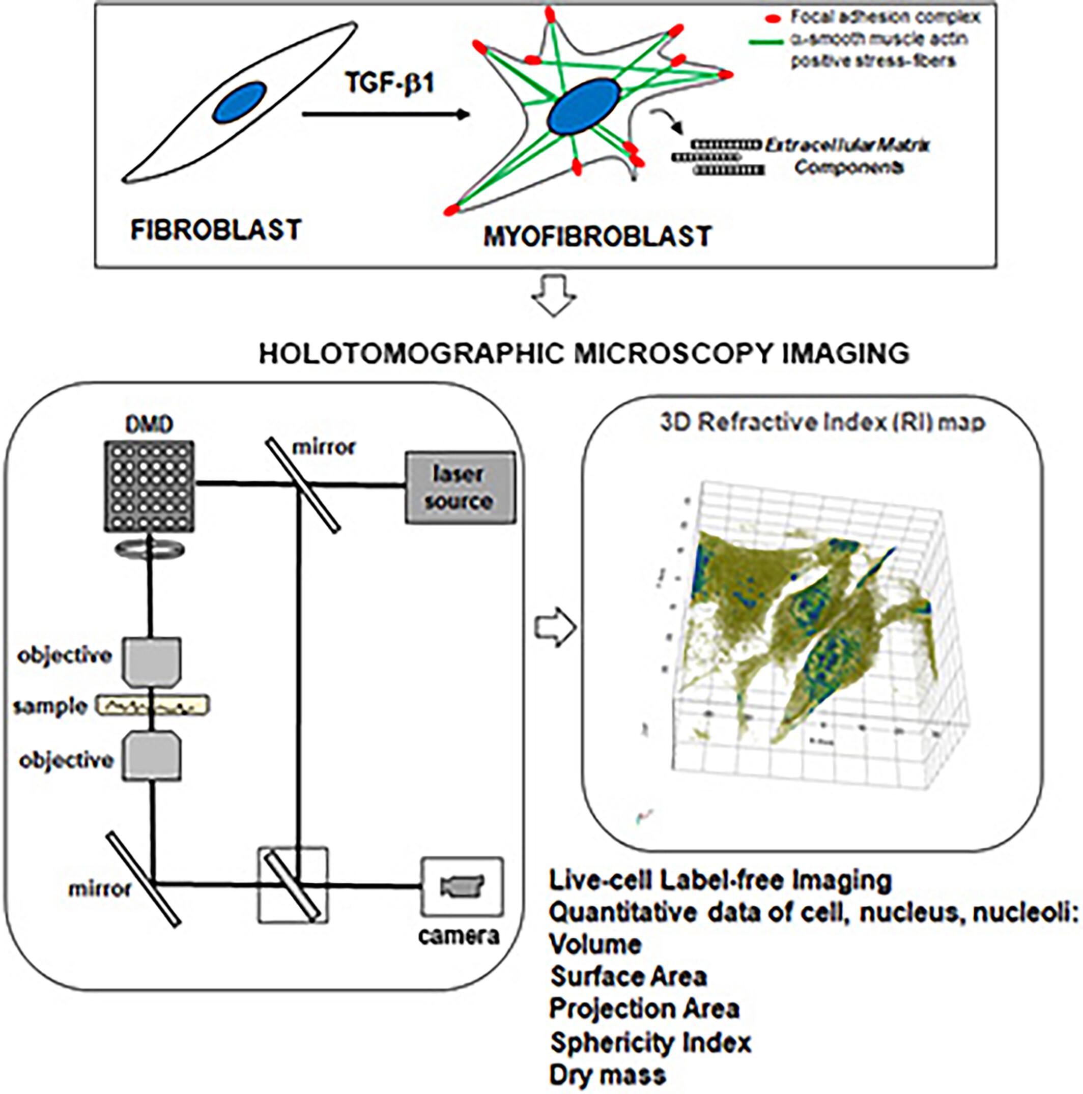
Holotomography (HT) is a cutting-edge fast live-cell quantitative label-free imaging technique. Based on the principle of quantitative phase imaging, it combines holography and tomography to record a three-dimensional map of the refractive index, used as intrinsic optical and quantitative imaging contrast parameter of biological samples, at a sub-micrometer spatial resolution. In this study HT has been employed for the first time to analyze the changes of fibroblasts differentiating towards myofibroblasts – recognized as the main cell player of fibrosis – when cultured in vitro with the pro-fibrotic factor, namely transforming growth factor-β1. In parallel, F-actin, vinculin, α-smooth muscle actin, phospho-myosin light chain 2, type-1 collagen, peroxisome proliferator-activated receptor-gamma coactivator-1α expression and mitochondria were evaluated by confocal laser scanning microscopy. Plasmamembrane passive properties and transient receptor potential canonical channels' currents were also recorded by whole-cell patch-clamp. The fluorescence images and electrophysiological results have been compared to the data obtained by HT and their congruence has been discussed. HT turned out to be a valid approach to morphologically distinguish fibroblasts from well differentiated myofibroblasts while obtaining objective measures concerning volume, surface area, projection area, surface index and dry mass (i.e., the mass of the non-aqueous content inside the cell including proteins and subcellular organelles) of the entire cell, nuclei and nucleoli with the major advantage to monitor outer and inner features in living cells in a non-invasive, rapid and label-free approach. HT might open up new research opportunities in the field of fibrotic diseases.
Research Highlights
Holotomography (HT) is a label-free laser interferometric imaging technology exploiting the intrinsic optical property of cells namely refractive index (RI) to enable a direct imaging and analysis of whole cells or intracellular organelles.
HT turned out a valid approach to distinguish morphological features of living unlabeled fibroblasts from differentiated myofibroblasts.
HT provided quantitative information concerning volume, surface area, projection area, surface index and dry mass of the entire fibroblasts/myofibroblasts, nuclei and nucleoli.
期刊介绍:
Microscopy Research and Technique (MRT) publishes articles on all aspects of advanced microscopy original architecture and methodologies with applications in the biological, clinical, chemical, and materials sciences. Original basic and applied research as well as technical papers dealing with the various subsets of microscopy are encouraged. MRT is the right form for those developing new microscopy methods or using the microscope to answer key questions in basic and applied research.


 分享
分享
 求助内容:
求助内容: 应助结果提醒方式:
 扫码关注我们
