{"title":"Characterization of Maximum Wall Shear Stress Points in Unruptured Cerebral Aneurysms Using Four-dimensional Flow Magnetic Resonance Imaging.","authors":"Kazuya Futami, Kouichi Misaki, Takehiro Uno, Iku Nambu, Tomoya Kamide, Mitsutoshi Nakada","doi":"10.1007/s00062-024-01436-w","DOIUrl":null,"url":null,"abstract":"<p><strong>Background: </strong>Maximum wall shear stress (maxWSS) points of unruptured cerebral aneurysms (UCAs) may cause wall remodeling leading to rupture. We characterized maxWSS points and their inherent intra-aneurysmal flow structures in a sizable cohort of saccular UCAs using four-dimensional (4D) flow magnetic resonance imaging (MRI).</p><p><strong>Methods: </strong>After contrast administration, 50 saccular UCAs were subjected to 4D flow MRI using a 1.5 T MRI scanner. Post-processing of obtained data was performed using commercially available software. The maxWSS points and maxWSS values were evaluated. The maxWSS values were statistically compared between aneurysm groups.</p><p><strong>Results: </strong>The maxWSS point was located on the aneurysm apex in 9 (18.0%), body in 2 (4.0%), and neck in 39 (78.0%) UCAs. The inherent intra-aneurysmal flow structure of the maxWSS point was an inflow zone in 34 (68.0%) UCAs, an inflow jet in 8 (16.0%), and an impingement zone in 8 (16.0%). The maxWSS point on the neck had significantly higher maxWSS values than those points on the other wall areas (P = 0.008). The maxWSS values of the maxWSS points on the apex and on the impingement zone were not significantly different compared with those of the other maxWSS points.</p><p><strong>Conclusion: </strong>The maxWSS points existed preferentially on the aneurysmal neck adjacent to the inflow zone with higher maxWSS values. The maxWSS points existed occasionally on the aneurysmal apex adjacent to the impingement zone. 4D flow MRI may be helpful to discriminate saccular UCAs with higher-risk maxWSS points that can cause wall remodeling leading to rupture.</p>","PeriodicalId":49298,"journal":{"name":"Clinical Neuroradiology","volume":" ","pages":"899-906"},"PeriodicalIF":3.2000,"publicationDate":"2024-12-01","publicationTypes":"Journal Article","fieldsOfStudy":null,"isOpenAccess":false,"openAccessPdf":"","citationCount":"0","resultStr":null,"platform":"Semanticscholar","paperid":null,"PeriodicalName":"Clinical Neuroradiology","FirstCategoryId":"1085","ListUrlMain":"https://doi.org/10.1007/s00062-024-01436-w","RegionNum":3,"RegionCategory":"医学","ArticlePicture":[],"TitleCN":null,"AbstractTextCN":null,"PMCID":null,"EPubDate":"2024/7/17 0:00:00","PubModel":"Epub","JCR":"Q2","JCRName":"CLINICAL NEUROLOGY","Score":null,"Total":0}
引用次数: 0
Abstract
Background: Maximum wall shear stress (maxWSS) points of unruptured cerebral aneurysms (UCAs) may cause wall remodeling leading to rupture. We characterized maxWSS points and their inherent intra-aneurysmal flow structures in a sizable cohort of saccular UCAs using four-dimensional (4D) flow magnetic resonance imaging (MRI).
Methods: After contrast administration, 50 saccular UCAs were subjected to 4D flow MRI using a 1.5 T MRI scanner. Post-processing of obtained data was performed using commercially available software. The maxWSS points and maxWSS values were evaluated. The maxWSS values were statistically compared between aneurysm groups.
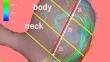
Results: The maxWSS point was located on the aneurysm apex in 9 (18.0%), body in 2 (4.0%), and neck in 39 (78.0%) UCAs. The inherent intra-aneurysmal flow structure of the maxWSS point was an inflow zone in 34 (68.0%) UCAs, an inflow jet in 8 (16.0%), and an impingement zone in 8 (16.0%). The maxWSS point on the neck had significantly higher maxWSS values than those points on the other wall areas (P = 0.008). The maxWSS values of the maxWSS points on the apex and on the impingement zone were not significantly different compared with those of the other maxWSS points.
Conclusion: The maxWSS points existed preferentially on the aneurysmal neck adjacent to the inflow zone with higher maxWSS values. The maxWSS points existed occasionally on the aneurysmal apex adjacent to the impingement zone. 4D flow MRI may be helpful to discriminate saccular UCAs with higher-risk maxWSS points that can cause wall remodeling leading to rupture.
期刊介绍:
Clinical Neuroradiology provides current information, original contributions, and reviews in the field of neuroradiology. An interdisciplinary approach is accomplished by diagnostic and therapeutic contributions related to associated subjects.
The international coverage and relevance of the journal is underlined by its being the official journal of the German, Swiss, and Austrian Societies of Neuroradiology.


 分享
分享
 求助内容:
求助内容: 应助结果提醒方式:
 扫码关注我们
