C T Arendt, C Uckermark, L Kovacheva, F Lieschke, R Golbach, S Edwin Thanarajah, E Hattingen, S Weidauer
{"title":"Wernicke Encephalopathy: Typical and Atypical Findings in Alcoholics and Non-Alcoholics and Correlation with Clinical Symptoms.","authors":"C T Arendt, C Uckermark, L Kovacheva, F Lieschke, R Golbach, S Edwin Thanarajah, E Hattingen, S Weidauer","doi":"10.1007/s00062-024-01434-y","DOIUrl":null,"url":null,"abstract":"<p><strong>Purpose: </strong>Clinical diagnosis of Wernicke encephalopathy (WE) can be challenging due to incomplete presentation of the classical triad. The aim was to provide an update on the relevance of standard MRI and to put typical and atypical imaging findings into context with clinical features.</p><p><strong>Methods: </strong>In this two-center retrospective observational study, the local radiology information system was searched for consecutive patients with clinical or imaging suspicion of WE. Two independent raters evaluated T2-weighted imaging (WI), fluid-attenuation inversion recovery (FLAIR), diffusion WI (DWI), T2*WI and/or susceptibility WI (SWI), and contrast-enhanced (CE)-T1WI, and noted the involvement of typical (i.e., mammillary bodies (MB), periaqueductal grey (PAG), thalamus, hypothalamus, tectal plate) and atypical (all others) lesion sites. Unusual signal patterns like hemorrhages were also documented. Reported clinical features together with the diagnostic criteria of the latest guidelines of the European Federation of Neurological Societies (EFNS) were used to test for relationships with MRI biomarkers.</p><p><strong>Results: </strong>47 patients with clinically confirmed WE were included (Jan '99-Apr '23; mean age, 53 yrs; 70% males). Interrater reliability for imaging findings was substantial (κ = 0.71), with lowest agreements for T2WI (κ = 0.85) compared to all other sequences and for PAG (κ = 0.65) compared to all other typical regions. In consensus, 77% (n = 36/47) of WE cases were rated MRI positive, with FLAIR (n = 36/47, 77%) showing the strongest relation (χ<sup>2</sup> = 47.0; P < 0.001) compared to all other sequences. Microbleeds in the MB were detected in four out of ten patients who received SWI, not visible on corresponding T2*WI. Atypical findings were observed in 23% (n = 11/47) of cases, always alongside typical findings, in both alcoholics (n = 9/44, 21%) and non-alcoholics (n = 2/3, 67%). Isolated involvement of structures, explicitly PAG (n = 4/36; 11%) or MB (n = 1/36; 3%), was present but observed less frequently than combined lesions (n = 31/36; 86%). A cut-off width of 2.5 mm for the PAG on 2D axial FLAIR was established between cases and age- and sex-matched controls. An independent association was demonstrated only between short-term memory loss and changes in the MB (OR = 2.2 [95% CI: 1.1-4.5]; P = 0.024). In retrospect, EFNS criteria were positive (≥ 2 out of 4) in every case, but its count (range, 2-4) showed no significant (P = 0.427) relationship with signal changes on standard MRI.</p><p><strong>Conclusion: </strong>The proposed sequence protocol (FLAIR, DWI, SWI and T1WI + CE) yielded good detection rates for neuroradiological findings in WE, with SWI showing microbleeds in the MB with superior detectability. However, false negative results in about a quarter of cases underline the importance of neurological alertness for the diagnosis. Awareness of atypical MRI findings should be raised, not only in non-alcoholics. There is limited correlation between clinical signs and standard MRI biomarkers.</p>","PeriodicalId":49298,"journal":{"name":"Clinical Neuroradiology","volume":" ","pages":"881-897"},"PeriodicalIF":2.6000,"publicationDate":"2024-12-01","publicationTypes":"Journal Article","fieldsOfStudy":null,"isOpenAccess":false,"openAccessPdf":"","citationCount":"0","resultStr":null,"platform":"Semanticscholar","paperid":null,"PeriodicalName":"Clinical Neuroradiology","FirstCategoryId":"3","ListUrlMain":"https://doi.org/10.1007/s00062-024-01434-y","RegionNum":3,"RegionCategory":"医学","ArticlePicture":[],"TitleCN":null,"AbstractTextCN":null,"PMCID":null,"EPubDate":"2024/7/16 0:00:00","PubModel":"Epub","JCR":"Q2","JCRName":"CLINICAL NEUROLOGY","Score":null,"Total":0}
引用次数: 0
Abstract
Purpose: Clinical diagnosis of Wernicke encephalopathy (WE) can be challenging due to incomplete presentation of the classical triad. The aim was to provide an update on the relevance of standard MRI and to put typical and atypical imaging findings into context with clinical features.
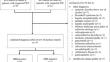
Methods: In this two-center retrospective observational study, the local radiology information system was searched for consecutive patients with clinical or imaging suspicion of WE. Two independent raters evaluated T2-weighted imaging (WI), fluid-attenuation inversion recovery (FLAIR), diffusion WI (DWI), T2*WI and/or susceptibility WI (SWI), and contrast-enhanced (CE)-T1WI, and noted the involvement of typical (i.e., mammillary bodies (MB), periaqueductal grey (PAG), thalamus, hypothalamus, tectal plate) and atypical (all others) lesion sites. Unusual signal patterns like hemorrhages were also documented. Reported clinical features together with the diagnostic criteria of the latest guidelines of the European Federation of Neurological Societies (EFNS) were used to test for relationships with MRI biomarkers.
Results: 47 patients with clinically confirmed WE were included (Jan '99-Apr '23; mean age, 53 yrs; 70% males). Interrater reliability for imaging findings was substantial (κ = 0.71), with lowest agreements for T2WI (κ = 0.85) compared to all other sequences and for PAG (κ = 0.65) compared to all other typical regions. In consensus, 77% (n = 36/47) of WE cases were rated MRI positive, with FLAIR (n = 36/47, 77%) showing the strongest relation (χ2 = 47.0; P < 0.001) compared to all other sequences. Microbleeds in the MB were detected in four out of ten patients who received SWI, not visible on corresponding T2*WI. Atypical findings were observed in 23% (n = 11/47) of cases, always alongside typical findings, in both alcoholics (n = 9/44, 21%) and non-alcoholics (n = 2/3, 67%). Isolated involvement of structures, explicitly PAG (n = 4/36; 11%) or MB (n = 1/36; 3%), was present but observed less frequently than combined lesions (n = 31/36; 86%). A cut-off width of 2.5 mm for the PAG on 2D axial FLAIR was established between cases and age- and sex-matched controls. An independent association was demonstrated only between short-term memory loss and changes in the MB (OR = 2.2 [95% CI: 1.1-4.5]; P = 0.024). In retrospect, EFNS criteria were positive (≥ 2 out of 4) in every case, but its count (range, 2-4) showed no significant (P = 0.427) relationship with signal changes on standard MRI.
Conclusion: The proposed sequence protocol (FLAIR, DWI, SWI and T1WI + CE) yielded good detection rates for neuroradiological findings in WE, with SWI showing microbleeds in the MB with superior detectability. However, false negative results in about a quarter of cases underline the importance of neurological alertness for the diagnosis. Awareness of atypical MRI findings should be raised, not only in non-alcoholics. There is limited correlation between clinical signs and standard MRI biomarkers.
期刊介绍:
Clinical Neuroradiology provides current information, original contributions, and reviews in the field of neuroradiology. An interdisciplinary approach is accomplished by diagnostic and therapeutic contributions related to associated subjects.
The international coverage and relevance of the journal is underlined by its being the official journal of the German, Swiss, and Austrian Societies of Neuroradiology.


 分享
分享
 求助内容:
求助内容: 应助结果提醒方式:
 扫码关注我们
