You-Jin Choi, Hye-Won Hu, Soo-Bin Kim, Ji-Hyun Lee, Seong-Taek Kim, Hee-Jin Kim
{"title":"Sihler's staining of the anterior belly of digastric muscle for botulinum toxin injection.","authors":"You-Jin Choi, Hye-Won Hu, Soo-Bin Kim, Ji-Hyun Lee, Seong-Taek Kim, Hee-Jin Kim","doi":"10.1007/s00276-024-03440-8","DOIUrl":null,"url":null,"abstract":"<p><strong>Purpose: </strong>The anterior belly of the digastric muscle (ABDM) is the target of botulinum toxin injection; however, anatomical considerations related to the injection point are absent. This study used Sihler's staining to analyze the intramuscular nerve distribution of ABDM to identify the most effective botulinum toxin injection points.</p><p><strong>Methods: </strong>We used 12 specimens from 6 embalmed cadavers in this study. The specimens were manually dissected to preserve the mylohyoid nerve and subjected to Sihler's staining. From the gnathion to and hyoid bone, the ABDM was divided into three equal parts, distinguishing the anterior, middle, and posterior thirds.</p><p><strong>Results: </strong>Only a branch of the mylohyoid nerve entered the ABDM, and its entry point was located in the middle-third region in all cases. The nerve endings were concentrated in the middle third (100%), followed by the anterior third (58.3%) and were not observed in the posterior third.</p><p><strong>Conclusion: </strong>The landmarks used in this study (gnathion and hyoid bone) are easily palpable on the skin surface, allowing clinicians to target the most effective injection site (middle third of ABDM). These results provide scientific and anatomic evidence for injection points, and will aid in the management of ABDM injection procedures in clinical practice.</p>","PeriodicalId":49461,"journal":{"name":"Surgical and Radiologic Anatomy","volume":" ","pages":"1543-1548"},"PeriodicalIF":1.2000,"publicationDate":"2024-09-01","publicationTypes":"Journal Article","fieldsOfStudy":null,"isOpenAccess":false,"openAccessPdf":"","citationCount":"0","resultStr":null,"platform":"Semanticscholar","paperid":null,"PeriodicalName":"Surgical and Radiologic Anatomy","FirstCategoryId":"3","ListUrlMain":"https://doi.org/10.1007/s00276-024-03440-8","RegionNum":4,"RegionCategory":"医学","ArticlePicture":[],"TitleCN":null,"AbstractTextCN":null,"PMCID":null,"EPubDate":"2024/7/16 0:00:00","PubModel":"Epub","JCR":"Q2","JCRName":"Medicine","Score":null,"Total":0}
引用次数: 0
Abstract
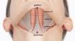
Purpose: The anterior belly of the digastric muscle (ABDM) is the target of botulinum toxin injection; however, anatomical considerations related to the injection point are absent. This study used Sihler's staining to analyze the intramuscular nerve distribution of ABDM to identify the most effective botulinum toxin injection points.
Methods: We used 12 specimens from 6 embalmed cadavers in this study. The specimens were manually dissected to preserve the mylohyoid nerve and subjected to Sihler's staining. From the gnathion to and hyoid bone, the ABDM was divided into three equal parts, distinguishing the anterior, middle, and posterior thirds.
Results: Only a branch of the mylohyoid nerve entered the ABDM, and its entry point was located in the middle-third region in all cases. The nerve endings were concentrated in the middle third (100%), followed by the anterior third (58.3%) and were not observed in the posterior third.
Conclusion: The landmarks used in this study (gnathion and hyoid bone) are easily palpable on the skin surface, allowing clinicians to target the most effective injection site (middle third of ABDM). These results provide scientific and anatomic evidence for injection points, and will aid in the management of ABDM injection procedures in clinical practice.
期刊介绍:
Anatomy is a morphological science which cannot fail to interest the clinician. The practical application of anatomical research to clinical problems necessitates special adaptation and selectivity in choosing from numerous international works. Although there is a tendency to believe that meaningful advances in anatomy are unlikely, constant revision is necessary. Surgical and Radiologic Anatomy, the first international journal of Clinical anatomy has been created in this spirit.
Its goal is to serve clinicians, regardless of speciality-physicians, surgeons, radiologists or other specialists-as an indispensable aid with which they can improve their knowledge of anatomy. Each issue includes: Original papers, review articles, articles on the anatomical bases of medical, surgical and radiological techniques, articles of normal radiologic anatomy, brief reviews of anatomical publications of clinical interest.
Particular attention is given to high quality illustrations, which are indispensable for a better understanding of anatomical problems.
Surgical and Radiologic Anatomy is a journal written by anatomists for clinicians with a special interest in anatomy.


 分享
分享
 求助内容:
求助内容: 应助结果提醒方式:
 扫码关注我们
