Laura P Hale, Andrew N Macintyre, Dawn E Bowles, Jean Kwun, Jie Li, Barbara Theriot, Joseph W Turek
{"title":"Comprehensive Flow Cytometric, Immunohistologic, and Molecular Assessment of Thymus Function in Rhesus Macaques.","authors":"Laura P Hale, Andrew N Macintyre, Dawn E Bowles, Jean Kwun, Jie Li, Barbara Theriot, Joseph W Turek","doi":"10.4049/immunohorizons.2300112","DOIUrl":null,"url":null,"abstract":"<p><p>The critical importance of the thymus for generating new naive T cells that protect against novel infections and are tolerant to self-antigens has led to a recent revival of interest in monitoring thymic function in species other than humans and mice. Nonhuman primates such as rhesus macaques (Macaca mulatta) provide particularly useful animal models for translational research in immunology. In this study, we tested the performance of a 15-marker multicolor Ab panel for flow cytometric phenotyping of lymphocyte subsets directly from rhesus whole blood, with validation by thymectomy and T cell depletion. Immunohistochemical and multiplex RNA expression analysis of thymus tissue biopsies and molecular assays on PBMCs were used to further validate thymus function. Results identify Ab panels that can accurately classify rhesus naive T cells (CD3+CD45RA+CD197+ or CD3+CD28+CD95-) and recent thymic emigrants (CD8+CD28+CD95-CD103+CD197+) using just 100 µl of whole blood and commercially available fluorescent Abs. An immunohistochemical panel reactive with pan-cytokeratin (CK), CK14, CD3, Ki-67, CCL21, and TdT provides histologic evidence of thymopoiesis from formalin-fixed, paraffin-embedded thymus tissues. Identification of mRNAs characteristic of both functioning thymic epithelial cells and developing thymocytes and/or molecular detection of products of TCR gene rearrangement provide additional complementary methods to evaluate thymopoiesis, without requiring specific Abs. Combinations of multiparameter flow cytometry, immunohistochemistry, multiplex gene expression, and TCR excision circle assays can comprehensively evaluate thymus function in rhesus macaques while requiring only minimal amounts of peripheral blood or biopsied thymus tissue.</p>","PeriodicalId":94037,"journal":{"name":"ImmunoHorizons","volume":"8 7","pages":"500-510"},"PeriodicalIF":0.0000,"publicationDate":"2024-07-01","publicationTypes":"Journal Article","fieldsOfStudy":null,"isOpenAccess":false,"openAccessPdf":"https://www.ncbi.nlm.nih.gov/pmc/articles/PMC11294275/pdf/","citationCount":"0","resultStr":null,"platform":"Semanticscholar","paperid":null,"PeriodicalName":"ImmunoHorizons","FirstCategoryId":"1085","ListUrlMain":"https://doi.org/10.4049/immunohorizons.2300112","RegionNum":0,"RegionCategory":null,"ArticlePicture":[],"TitleCN":null,"AbstractTextCN":null,"PMCID":null,"EPubDate":"","PubModel":"","JCR":"Q3","JCRName":"Medicine","Score":null,"Total":0}
引用次数: 0
Abstract
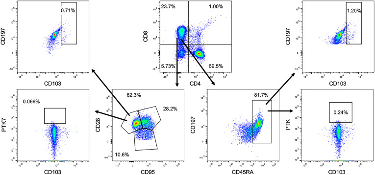
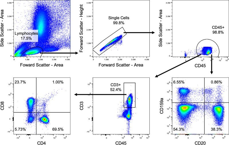
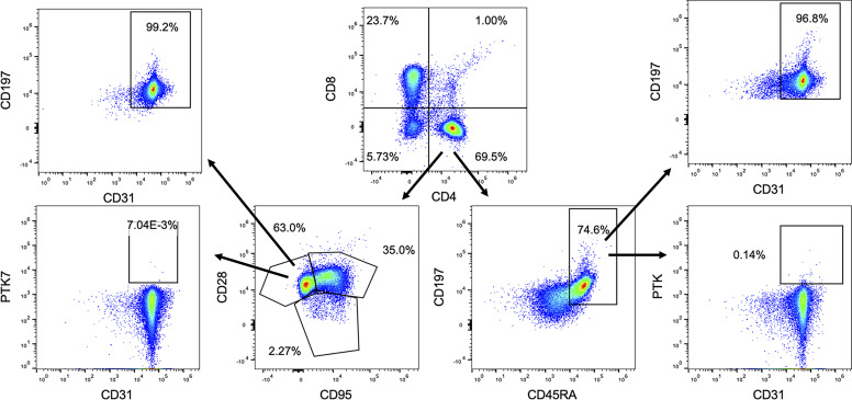
The critical importance of the thymus for generating new naive T cells that protect against novel infections and are tolerant to self-antigens has led to a recent revival of interest in monitoring thymic function in species other than humans and mice. Nonhuman primates such as rhesus macaques (Macaca mulatta) provide particularly useful animal models for translational research in immunology. In this study, we tested the performance of a 15-marker multicolor Ab panel for flow cytometric phenotyping of lymphocyte subsets directly from rhesus whole blood, with validation by thymectomy and T cell depletion. Immunohistochemical and multiplex RNA expression analysis of thymus tissue biopsies and molecular assays on PBMCs were used to further validate thymus function. Results identify Ab panels that can accurately classify rhesus naive T cells (CD3+CD45RA+CD197+ or CD3+CD28+CD95-) and recent thymic emigrants (CD8+CD28+CD95-CD103+CD197+) using just 100 µl of whole blood and commercially available fluorescent Abs. An immunohistochemical panel reactive with pan-cytokeratin (CK), CK14, CD3, Ki-67, CCL21, and TdT provides histologic evidence of thymopoiesis from formalin-fixed, paraffin-embedded thymus tissues. Identification of mRNAs characteristic of both functioning thymic epithelial cells and developing thymocytes and/or molecular detection of products of TCR gene rearrangement provide additional complementary methods to evaluate thymopoiesis, without requiring specific Abs. Combinations of multiparameter flow cytometry, immunohistochemistry, multiplex gene expression, and TCR excision circle assays can comprehensively evaluate thymus function in rhesus macaques while requiring only minimal amounts of peripheral blood or biopsied thymus tissue.


 分享
分享

 求助内容:
求助内容: 应助结果提醒方式:
 扫码关注我们
