Jiaqi Tan , Chu Zhang , Ziyi Bao , Hanyang Zhao , Li Zhang , Hongxi Xu
{"title":"A new insight into the mechanism of dichlorodiphenyltrichloroethane-induced hepatotoxicity based on GSDME-mediated pyroptosis","authors":"Jiaqi Tan , Chu Zhang , Ziyi Bao , Hanyang Zhao , Li Zhang , Hongxi Xu","doi":"10.1016/j.pestbp.2024.106030","DOIUrl":null,"url":null,"abstract":"<div><p>There have been persistent concerns about the safety risks associated with DDT residues in the environment. Studies have shown that exposure to DDT or its metabolites can cause various liver diseases. However, the mechanisms of liver toxicity haven't been well studied. In our current investigation, we observed that DDT triggers pyroptosis in human liver cells (HL-7702), representing a novel form of programmed cell death. Our results delineated DDT (0–100 μM) induced pyroptosis in HL-7702 cells, which was confirmed through morphological changes, lactate dehydrogenase (LDH) release, gasdermin E (GSDME) cleavage and Annexin-V/PI staining. Knockdown of GSDME reduced cell death and transferred the mode of cell death from pyroptosis to apoptosis. Notably, DDT exposure markedly increased reactive oxygen species (ROS) production, concurrent with c-Jun N-terminal kinase (JNK) phosphorylation. Intervention with a ROS inhibitor or JNK inhibitor SP600125 restored cell viability and hindered GSDME-mediated pyroptosis. Our results firstly demonstrate that DDT suppresses HL-7702 cells growth by inducing pyroptosis mainly through the ROS/JNK/GSDME pathway. These findings not only contribute to an in-depth understanding of DDT toxicity but also open avenues for gaining valuable insights into potential mitigation strategies and therapeutic interventions.</p></div>","PeriodicalId":19828,"journal":{"name":"Pesticide Biochemistry and Physiology","volume":"204 ","pages":"Article 106030"},"PeriodicalIF":4.0000,"publicationDate":"2024-09-01","publicationTypes":"Journal Article","fieldsOfStudy":null,"isOpenAccess":false,"openAccessPdf":"","citationCount":"0","resultStr":null,"platform":"Semanticscholar","paperid":null,"PeriodicalName":"Pesticide Biochemistry and Physiology","FirstCategoryId":"97","ListUrlMain":"https://www.sciencedirect.com/science/article/pii/S0048357524002633","RegionNum":1,"RegionCategory":"农林科学","ArticlePicture":[],"TitleCN":null,"AbstractTextCN":null,"PMCID":null,"EPubDate":"2024/7/15 0:00:00","PubModel":"Epub","JCR":"Q2","JCRName":"BIOCHEMISTRY & MOLECULAR BIOLOGY","Score":null,"Total":0}
引用次数: 0
Abstract
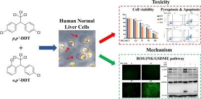
There have been persistent concerns about the safety risks associated with DDT residues in the environment. Studies have shown that exposure to DDT or its metabolites can cause various liver diseases. However, the mechanisms of liver toxicity haven't been well studied. In our current investigation, we observed that DDT triggers pyroptosis in human liver cells (HL-7702), representing a novel form of programmed cell death. Our results delineated DDT (0–100 μM) induced pyroptosis in HL-7702 cells, which was confirmed through morphological changes, lactate dehydrogenase (LDH) release, gasdermin E (GSDME) cleavage and Annexin-V/PI staining. Knockdown of GSDME reduced cell death and transferred the mode of cell death from pyroptosis to apoptosis. Notably, DDT exposure markedly increased reactive oxygen species (ROS) production, concurrent with c-Jun N-terminal kinase (JNK) phosphorylation. Intervention with a ROS inhibitor or JNK inhibitor SP600125 restored cell viability and hindered GSDME-mediated pyroptosis. Our results firstly demonstrate that DDT suppresses HL-7702 cells growth by inducing pyroptosis mainly through the ROS/JNK/GSDME pathway. These findings not only contribute to an in-depth understanding of DDT toxicity but also open avenues for gaining valuable insights into potential mitigation strategies and therapeutic interventions.
期刊介绍:
Pesticide Biochemistry and Physiology publishes original scientific articles pertaining to the mode of action of plant protection agents such as insecticides, fungicides, herbicides, and similar compounds, including nonlethal pest control agents, biosynthesis of pheromones, hormones, and plant resistance agents. Manuscripts may include a biochemical, physiological, or molecular study for an understanding of comparative toxicology or selective toxicity of both target and nontarget organisms. Particular interest will be given to studies on the molecular biology of pest control, toxicology, and pesticide resistance.
Research Areas Emphasized Include the Biochemistry and Physiology of:
• Comparative toxicity
• Mode of action
• Pathophysiology
• Plant growth regulators
• Resistance
• Other effects of pesticides on both parasites and hosts.


 分享
分享
 求助内容:
求助内容: 应助结果提醒方式:
 扫码关注我们
