Huili Wang, Jianfeng Qiu, Weizhao Lu, Jindong Xie, Junchi Ma
{"title":"Radiomics based on multiple machine learning methods for diagnosing early bone metastases not visible on CT images.","authors":"Huili Wang, Jianfeng Qiu, Weizhao Lu, Jindong Xie, Junchi Ma","doi":"10.1007/s00256-024-04752-x","DOIUrl":null,"url":null,"abstract":"<p><strong>Objectives: </strong>This study utilizes [<sup>99m</sup>Tc]-methylene diphosphate (MDP) single photon emission computed tomography (SPECT) images as a reference standard to evaluate whether the integration of radiomics features from computed tomography (CT) and machine learning algorithms can identify microscopic early bone metastases. Additionally, we also determine the optimal machine learning approach.</p><p><strong>Materials and methods: </strong>We retrospectively studied 63 patients with early bone metastasis from July 2020 to March 2023. The ITK-SNAP software was used to delineate early bone metastases and normal bone tissue in SPECT images of each patient, which were then registered onto CT images to outline the volume of interest (VOI). The VOI includes 63 early bone metastasis volumes and 63 normal bone tissue volumes. 126 VOIs were randomly distributed in a 7:3 ratio between the training and testing groups, and 944 radiomics features were extracted from every VOI. We established 20 machine learning models using 5 feature selection algorithms and 4 classification methods. Evaluate the performance of the model using the area under the receiver operating characteristic curve (AUC).</p><p><strong>Results: </strong>Most machine learning models demonstrated outstanding discriminative capacity, with AUCs higher than 0.70. Notably, the K-Nearest Neighbors (KNN) classifier exhibited significant performance improvement compared to the other four classifiers. Specifically, the model constructed utilizing eXtreme Gradient Boosting (XGBoost) feature selection method integrated with KNN classifier achieved the maximum AUC, which is 0.989 in the training set and 0.975 in the testing set.</p><p><strong>Conclusions: </strong>Radiomics features integrated with machine learning methods can identify early bone metastases that are not visible on CT images. In our analysis, KNN is considered the optimal classification method.</p>","PeriodicalId":21783,"journal":{"name":"Skeletal Radiology","volume":" ","pages":"335-343"},"PeriodicalIF":2.2000,"publicationDate":"2025-02-01","publicationTypes":"Journal Article","fieldsOfStudy":null,"isOpenAccess":false,"openAccessPdf":"","citationCount":"0","resultStr":null,"platform":"Semanticscholar","paperid":null,"PeriodicalName":"Skeletal Radiology","FirstCategoryId":"3","ListUrlMain":"https://doi.org/10.1007/s00256-024-04752-x","RegionNum":3,"RegionCategory":"医学","ArticlePicture":[],"TitleCN":null,"AbstractTextCN":null,"PMCID":null,"EPubDate":"2024/7/19 0:00:00","PubModel":"Epub","JCR":"Q2","JCRName":"ORTHOPEDICS","Score":null,"Total":0}
引用次数: 0
Abstract
Objectives: This study utilizes [99mTc]-methylene diphosphate (MDP) single photon emission computed tomography (SPECT) images as a reference standard to evaluate whether the integration of radiomics features from computed tomography (CT) and machine learning algorithms can identify microscopic early bone metastases. Additionally, we also determine the optimal machine learning approach.
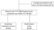
Materials and methods: We retrospectively studied 63 patients with early bone metastasis from July 2020 to March 2023. The ITK-SNAP software was used to delineate early bone metastases and normal bone tissue in SPECT images of each patient, which were then registered onto CT images to outline the volume of interest (VOI). The VOI includes 63 early bone metastasis volumes and 63 normal bone tissue volumes. 126 VOIs were randomly distributed in a 7:3 ratio between the training and testing groups, and 944 radiomics features were extracted from every VOI. We established 20 machine learning models using 5 feature selection algorithms and 4 classification methods. Evaluate the performance of the model using the area under the receiver operating characteristic curve (AUC).
Results: Most machine learning models demonstrated outstanding discriminative capacity, with AUCs higher than 0.70. Notably, the K-Nearest Neighbors (KNN) classifier exhibited significant performance improvement compared to the other four classifiers. Specifically, the model constructed utilizing eXtreme Gradient Boosting (XGBoost) feature selection method integrated with KNN classifier achieved the maximum AUC, which is 0.989 in the training set and 0.975 in the testing set.
Conclusions: Radiomics features integrated with machine learning methods can identify early bone metastases that are not visible on CT images. In our analysis, KNN is considered the optimal classification method.
期刊介绍:
Skeletal Radiology provides a forum for the dissemination of current knowledge and information dealing with disorders of the musculoskeletal system including the spine. While emphasizing the radiological aspects of the many varied skeletal abnormalities, the journal also adopts an interdisciplinary approach, reflecting the membership of the International Skeletal Society. Thus, the anatomical, pathological, physiological, clinical, metabolic and epidemiological aspects of the many entities affecting the skeleton receive appropriate consideration.
This is the Journal of the International Skeletal Society and the Official Journal of the Society of Skeletal Radiology and the Australasian Musculoskelelal Imaging Group.


 分享
分享
 求助内容:
求助内容: 应助结果提醒方式:
 扫码关注我们
