A cross-sectional CBCT assessment of the relative position of one-piece titanium-zirconium mini-implants placed for mandibular overdentures using non-guided surgery
Cláudio Rodrigues Leles, Leuçon de Oliveira Moura-Neto, Jésio Rodrigues Silva, Lays Noleto Nascimento, Thalita Fernandes Fleury Curado, Nadia Lago Costa, Martin Schimmel, Gerald McKenna
{"title":"A cross-sectional CBCT assessment of the relative position of one-piece titanium-zirconium mini-implants placed for mandibular overdentures using non-guided surgery","authors":"Cláudio Rodrigues Leles, Leuçon de Oliveira Moura-Neto, Jésio Rodrigues Silva, Lays Noleto Nascimento, Thalita Fernandes Fleury Curado, Nadia Lago Costa, Martin Schimmel, Gerald McKenna","doi":"10.1111/clr.14335","DOIUrl":null,"url":null,"abstract":"<div>\n \n \n <section>\n \n <h3> Objective</h3>\n \n <p>To assess the relative position of mini-implants to retain a mandibular overdenture, according to the surgical protocol, technical and anatomical factors.</p>\n </section>\n \n <section>\n \n <h3> Methods</h3>\n \n <p>Mandibular cone-beam computed tomography (CBCT) scans were analyzed for 73 patients who received four one-piece titanium-zirconium mini-implants. Drilling was performed using a 1.6 mm needle drill and a 2.2 mm Pilot Drill, according to the bone density with a surgical stent. Post-insertion CBCT images in DICOM format were analyzed using the E-Vol-DX software with BAR filters. Divergence angle between implants and between implants and the overdenture path of insertion was measured using CliniView 10.2.6 software.</p>\n </section>\n \n <section>\n \n <h3> Results</h3>\n \n <p>Divergence between implants ranged from 0° to 22.3° (mean = 4.2; SD = 3.7) in the lateral and from 0° to 26.2° (mean = 5.3; SD = 4.1) in the frontal projections (<i>p</i> < .001). Only 1 (0.2%) and 3 (0.7%) of the measurements were higher than 20° in the lateral and frontal views, respectively. The mean angulations between the implant and the path of insertion for the overdenture were 9.3° (SD = 7.5) and 4.0° (SD = 2.9) for the lateral and frontal views, respectively (<i>p</i> < .001). Regression analyses showed a significant association between the divergence of implants and the frontal view projection (<i>p</i> < .001), greater distance between the paired implants (<i>p</i> = .017), the flapped surgical protocol (<i>p</i> = .002), higher final insertion torque (<i>p</i> = .011), and deeper preparation with the needle drill (<i>p</i> < .001).</p>\n </section>\n \n <section>\n \n <h3> Conclusions</h3>\n \n <p>The mini-implants were placed with low divergence angles and satisfactory parallelism. Factors including shorter distances between the implants, higher density bone, and a flapless surgical approach all contributed positively to improved parallelism of the mini-implants.</p>\n </section>\n </div>","PeriodicalId":10455,"journal":{"name":"Clinical Oral Implants Research","volume":"35 11","pages":"1475-1484"},"PeriodicalIF":5.3000,"publicationDate":"2024-07-23","publicationTypes":"Journal Article","fieldsOfStudy":null,"isOpenAccess":false,"openAccessPdf":"https://onlinelibrary.wiley.com/doi/epdf/10.1111/clr.14335","citationCount":"0","resultStr":null,"platform":"Semanticscholar","paperid":null,"PeriodicalName":"Clinical Oral Implants Research","FirstCategoryId":"5","ListUrlMain":"https://onlinelibrary.wiley.com/doi/10.1111/clr.14335","RegionNum":1,"RegionCategory":"医学","ArticlePicture":[],"TitleCN":null,"AbstractTextCN":null,"PMCID":null,"EPubDate":"","PubModel":"","JCR":"Q1","JCRName":"DENTISTRY, ORAL SURGERY & MEDICINE","Score":null,"Total":0}
引用次数: 0
Abstract
Objective
To assess the relative position of mini-implants to retain a mandibular overdenture, according to the surgical protocol, technical and anatomical factors.
Methods
Mandibular cone-beam computed tomography (CBCT) scans were analyzed for 73 patients who received four one-piece titanium-zirconium mini-implants. Drilling was performed using a 1.6 mm needle drill and a 2.2 mm Pilot Drill, according to the bone density with a surgical stent. Post-insertion CBCT images in DICOM format were analyzed using the E-Vol-DX software with BAR filters. Divergence angle between implants and between implants and the overdenture path of insertion was measured using CliniView 10.2.6 software.
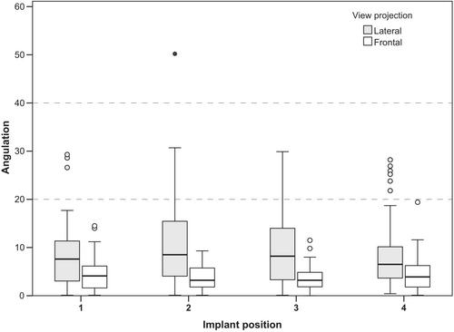
Results
Divergence between implants ranged from 0° to 22.3° (mean = 4.2; SD = 3.7) in the lateral and from 0° to 26.2° (mean = 5.3; SD = 4.1) in the frontal projections (p < .001). Only 1 (0.2%) and 3 (0.7%) of the measurements were higher than 20° in the lateral and frontal views, respectively. The mean angulations between the implant and the path of insertion for the overdenture were 9.3° (SD = 7.5) and 4.0° (SD = 2.9) for the lateral and frontal views, respectively (p < .001). Regression analyses showed a significant association between the divergence of implants and the frontal view projection (p < .001), greater distance between the paired implants (p = .017), the flapped surgical protocol (p = .002), higher final insertion torque (p = .011), and deeper preparation with the needle drill (p < .001).
Conclusions
The mini-implants were placed with low divergence angles and satisfactory parallelism. Factors including shorter distances between the implants, higher density bone, and a flapless surgical approach all contributed positively to improved parallelism of the mini-implants.
期刊介绍:
Clinical Oral Implants Research conveys scientific progress in the field of implant dentistry and its related areas to clinicians, teachers and researchers concerned with the application of this information for the benefit of patients in need of oral implants. The journal addresses itself to clinicians, general practitioners, periodontists, oral and maxillofacial surgeons and prosthodontists, as well as to teachers, academicians and scholars involved in the education of professionals and in the scientific promotion of the field of implant dentistry.


 分享
分享
 求助内容:
求助内容: 应助结果提醒方式:
 扫码关注我们
