Pae Sun Suh, Jung Hwan Baek, Jae Ho Lee, Sae Rom Chung, Young Jun Choi, Ki-Wook Chung, Tae Yong Kim, Jeong Hyun Lee
{"title":"Effectiveness of microvascular flow imaging for radiofrequency ablation in recurrent thyroid cancer: comparison with power Doppler imaging.","authors":"Pae Sun Suh, Jung Hwan Baek, Jae Ho Lee, Sae Rom Chung, Young Jun Choi, Ki-Wook Chung, Tae Yong Kim, Jeong Hyun Lee","doi":"10.1007/s00330-024-10977-0","DOIUrl":null,"url":null,"abstract":"<p><strong>Objectives: </strong>To compare microvascular flow imaging (MVFI) and power Doppler ultrasonography imaging (PDUS) for detecting intratumoral vascularity in recurrent thyroid cancer both before and after radiofrequency ablation (RFA).</p><p><strong>Methods: </strong>This retrospective study included 80 patients (age, 57 ± 12 years; 54 women) with 110 recurrent tumors who underwent RFA between January 2021 and June 2023. A total of 151 PDUS and MVFI image sets were analyzed (85 pre-RFA, 66 post-RFA). Two readers assessed vascularity on the images using a four-point scale with a 2-week interval between PDUS and MVFI to estimate inter-reader agreement. Intra-reader agreement was determined by reinterpreting images in reverse order (MVFI-PDUS) after a 1-month gap. Additionally, diagnostic performance for identifying viable tumors after RFA was assessed in 44 lesions using thyroid-protocol CT as a reference standard.</p><p><strong>Results: </strong>MVFI demonstrated higher vascular grades than PDUS, both before (reader 1: 3.04 ± 1.15 vs. 1.93 ± 1.07, p < 0.001; reader 2: 3.20 ± 0.96 vs. 2.12 ± 1.07, p < 0.001) and after RFA (reader 1: 2.44 ± 1.28 vs. 1.67 ± 1.06, p < 0.001; reader 2: 2.62 ± 1.23 vs. 1.83 ± 0.99, p < 0.001). Inter-reader agreement was substantial (κ = 0.743) and intra-reader agreement was almost perfect (κ = 0.840). MVFI showed higher sensitivity (81.5%-88.9%) and accuracy (84.1%-86.4%) than PDUS (sensitivity: 51.9%, p < 0.01; accuracy: 63.6-70.5%, p < 0.04), without sacrificing specificity.</p><p><strong>Conclusion: </strong>MVFI was superior to PDUS for assessing intratumoral vascularity and showed good inter- and intra-reader agreement, highlighting its clinical value for assessing pre-RFA vascularity and accurately identifying post-RFA viable tumors in recurrent thyroid cancer.</p><p><strong>Clinical relevance statement: </strong>Microvascular flow imaging (MVFI) is superior to power-Doppler US for assessing intratumoral vascularity; therefore, MVFI can be a valuable tool for assessing vascularity before radiofrequency ablation (RFA) and for identifying viable tumors after RFA in patients with recurrent thyroid cancer.</p><p><strong>Key points: </strong>The value of microvascular flow imaging (MVFI) for evaluating intratumoral vascularity is unexplored. MVFI demonstrated higher vascular grades than power Doppler US before and after ablation. Microvascular flow imaging showed higher sensitivity and accuracy than power Doppler US without sacrificing specificity.</p>","PeriodicalId":12076,"journal":{"name":"European Radiology","volume":" ","pages":"597-607"},"PeriodicalIF":4.7000,"publicationDate":"2025-02-01","publicationTypes":"Journal Article","fieldsOfStudy":null,"isOpenAccess":false,"openAccessPdf":"","citationCount":"0","resultStr":null,"platform":"Semanticscholar","paperid":null,"PeriodicalName":"European Radiology","FirstCategoryId":"3","ListUrlMain":"https://doi.org/10.1007/s00330-024-10977-0","RegionNum":2,"RegionCategory":"医学","ArticlePicture":[],"TitleCN":null,"AbstractTextCN":null,"PMCID":null,"EPubDate":"2024/7/23 0:00:00","PubModel":"Epub","JCR":"Q1","JCRName":"RADIOLOGY, NUCLEAR MEDICINE & MEDICAL IMAGING","Score":null,"Total":0}
引用次数: 0
Abstract
Objectives: To compare microvascular flow imaging (MVFI) and power Doppler ultrasonography imaging (PDUS) for detecting intratumoral vascularity in recurrent thyroid cancer both before and after radiofrequency ablation (RFA).
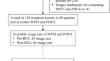
Methods: This retrospective study included 80 patients (age, 57 ± 12 years; 54 women) with 110 recurrent tumors who underwent RFA between January 2021 and June 2023. A total of 151 PDUS and MVFI image sets were analyzed (85 pre-RFA, 66 post-RFA). Two readers assessed vascularity on the images using a four-point scale with a 2-week interval between PDUS and MVFI to estimate inter-reader agreement. Intra-reader agreement was determined by reinterpreting images in reverse order (MVFI-PDUS) after a 1-month gap. Additionally, diagnostic performance for identifying viable tumors after RFA was assessed in 44 lesions using thyroid-protocol CT as a reference standard.
Results: MVFI demonstrated higher vascular grades than PDUS, both before (reader 1: 3.04 ± 1.15 vs. 1.93 ± 1.07, p < 0.001; reader 2: 3.20 ± 0.96 vs. 2.12 ± 1.07, p < 0.001) and after RFA (reader 1: 2.44 ± 1.28 vs. 1.67 ± 1.06, p < 0.001; reader 2: 2.62 ± 1.23 vs. 1.83 ± 0.99, p < 0.001). Inter-reader agreement was substantial (κ = 0.743) and intra-reader agreement was almost perfect (κ = 0.840). MVFI showed higher sensitivity (81.5%-88.9%) and accuracy (84.1%-86.4%) than PDUS (sensitivity: 51.9%, p < 0.01; accuracy: 63.6-70.5%, p < 0.04), without sacrificing specificity.
Conclusion: MVFI was superior to PDUS for assessing intratumoral vascularity and showed good inter- and intra-reader agreement, highlighting its clinical value for assessing pre-RFA vascularity and accurately identifying post-RFA viable tumors in recurrent thyroid cancer.
Clinical relevance statement: Microvascular flow imaging (MVFI) is superior to power-Doppler US for assessing intratumoral vascularity; therefore, MVFI can be a valuable tool for assessing vascularity before radiofrequency ablation (RFA) and for identifying viable tumors after RFA in patients with recurrent thyroid cancer.
Key points: The value of microvascular flow imaging (MVFI) for evaluating intratumoral vascularity is unexplored. MVFI demonstrated higher vascular grades than power Doppler US before and after ablation. Microvascular flow imaging showed higher sensitivity and accuracy than power Doppler US without sacrificing specificity.
期刊介绍:
European Radiology (ER) continuously updates scientific knowledge in radiology by publication of strong original articles and state-of-the-art reviews written by leading radiologists. A well balanced combination of review articles, original papers, short communications from European radiological congresses and information on society matters makes ER an indispensable source for current information in this field.
This is the Journal of the European Society of Radiology, and the official journal of a number of societies.
From 2004-2008 supplements to European Radiology were published under its companion, European Radiology Supplements, ISSN 1613-3749.


 分享
分享
 求助内容:
求助内容: 应助结果提醒方式:
 扫码关注我们
