{"title":"The fMRI global signal and its association with the signal from cranial bone","authors":"","doi":"10.1016/j.neuroimage.2024.120754","DOIUrl":null,"url":null,"abstract":"<div><p>The nature of the global signal, i.e. the average signal from sequential functional imaging scans of the brain or the cortex, is not well understood, but is thought to include vascular and neural components. Using resting state data, we report on the strong association between the global signal and the average signal from the part of the volume that includes the cranial bone and subdural vessels and venous collectors, separated from each other and the subdural space by multispectral segmentation procedures. While subdural vessels carried a signal with a phase delay relative to the cortex, the association with the cortical signal was strongest in the parts of the scan corresponding to the laminae of the cranial bone, reaching 80% shared variance in some individuals. These findings suggest that in resting state data vascular components may play a prominent role in the genesis of fluctuations of the global signal. Evidence from other studies on the existence of neural sources of the global signal suggests that it may reflect the action of multiple mechanisms (including cerebrovascular reactivity and autonomic control) concurrently acting to regulate global cerebral perfusion.</p></div>","PeriodicalId":19299,"journal":{"name":"NeuroImage","volume":null,"pages":null},"PeriodicalIF":4.7000,"publicationDate":"2024-07-25","publicationTypes":"Journal Article","fieldsOfStudy":null,"isOpenAccess":false,"openAccessPdf":"https://www.sciencedirect.com/science/article/pii/S1053811924002519/pdfft?md5=f1e47aa38d4b0dba8dcba80a45b566ad&pid=1-s2.0-S1053811924002519-main.pdf","citationCount":"0","resultStr":null,"platform":"Semanticscholar","paperid":null,"PeriodicalName":"NeuroImage","FirstCategoryId":"3","ListUrlMain":"https://www.sciencedirect.com/science/article/pii/S1053811924002519","RegionNum":2,"RegionCategory":"医学","ArticlePicture":[],"TitleCN":null,"AbstractTextCN":null,"PMCID":null,"EPubDate":"","PubModel":"","JCR":"Q1","JCRName":"NEUROIMAGING","Score":null,"Total":0}
引用次数: 0
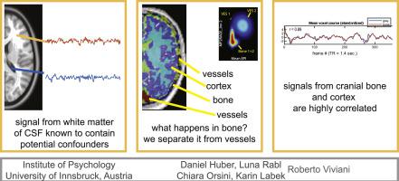
Abstract
The nature of the global signal, i.e. the average signal from sequential functional imaging scans of the brain or the cortex, is not well understood, but is thought to include vascular and neural components. Using resting state data, we report on the strong association between the global signal and the average signal from the part of the volume that includes the cranial bone and subdural vessels and venous collectors, separated from each other and the subdural space by multispectral segmentation procedures. While subdural vessels carried a signal with a phase delay relative to the cortex, the association with the cortical signal was strongest in the parts of the scan corresponding to the laminae of the cranial bone, reaching 80% shared variance in some individuals. These findings suggest that in resting state data vascular components may play a prominent role in the genesis of fluctuations of the global signal. Evidence from other studies on the existence of neural sources of the global signal suggests that it may reflect the action of multiple mechanisms (including cerebrovascular reactivity and autonomic control) concurrently acting to regulate global cerebral perfusion.
期刊介绍:
NeuroImage, a Journal of Brain Function provides a vehicle for communicating important advances in acquiring, analyzing, and modelling neuroimaging data and in applying these techniques to the study of structure-function and brain-behavior relationships. Though the emphasis is on the macroscopic level of human brain organization, meso-and microscopic neuroimaging across all species will be considered if informative for understanding the aforementioned relationships.


 分享
分享
 求助内容:
求助内容: 应助结果提醒方式:
 扫码关注我们
