{"title":"An SEM study on the effect of 9.3-µm CO<sub>2</sub> laser on dentinal tubules for hypersensitivity treatment.","authors":"Vijayashankar Ramareddy, Charles Kerbage","doi":"10.1007/s10103-024-04157-1","DOIUrl":null,"url":null,"abstract":"<p><strong>Objectives: </strong>In-vitro studies were performed on dentin of extracted human molars to investigate the effectiveness of 9.3 μm CO<sub>2</sub> laser irradiation to occlude dentinal tubules. The observed occlusion of dentinal tubules with the irradiation was compared with application of three reagents: 2% Sodium Fluoride gel, an aqueous solution of hydroxyapatite nanoparticles and an equal mix of the two. We show that 9.3 μm CO<sub>2</sub> laser irradiation occludes dentinal tubules, and the use of laser irradiation produces better occlusion of the opened tubules compared to the use of topical reagents.</p><p><strong>Methods: </strong>Nine extracted and cleaned human molars were cut to obtain dentin disks of thickness of 3-5 mm. Each disc was divided into four quarters, and each quarter served as two samples corresponding to irradiated and non-irradiated group counterparts. Five disks were used to study the effect of various laser irradiation energies on the dentinal tubules to find a good pulse fluence for occlusion of the dentinal tubules, and four disks were used for studying the effects of reagents and irradiation at the pulse fluences found in the first part of the study. The samples were irradiated with a beam diameter of 1 mm (1/e<sup>2</sup>) at 15 Hz pulse repetition rate, scanned automatically using a set of scanning mirrors. Samples were imaged using Scanning Electron Microscope (SEM) which were processed to determine tubule diameter. Safety of the irradiation treatment was investigated on 6 samples by measuring pulpal temperature rise. The effect of three topical reagents corresponding to 2% Sodium Fluoride gel (F), Hydroxyapatite nanoparticles (HA) and an equal mix of F and HA (HAF) on dentinal tubule occlusion was evaluated and compared with the laser irradiation.</p><p><strong>Results: </strong>In all examined cases, laser irradiation at a fluence of 0.81 J/cm<sup>2</sup> resulted in a temperature increase less than 3 °C which is safe, and no surface cracking was observed. There is a threshold pulse fluence of 0.27 J/cm<sup>2</sup> above which, laser produced surface melting. At a pulse fluence of 0.81 J/cm<sup>2</sup> a layer of recast of melted dentin was formed. Under this layer, peritubular dentin melting and occluding of the dentinal tubules was observed. Application of either F or HA or HAF did not produce visible occlusion effect on open tubules after washing and microbrushing with excess distilled water.</p><p><strong>Conclusions: </strong>9.3 μm CO2 laser irradiation on extracted human molar dentin at pulse fluence of 0.81 J./cm<sup>2</sup> resulted in tubule area reduction by 97% without rising pulpal temperatures to unsafe levels.</p>","PeriodicalId":17978,"journal":{"name":"Lasers in Medical Science","volume":"39 1","pages":"200"},"PeriodicalIF":2.4000,"publicationDate":"2024-07-31","publicationTypes":"Journal Article","fieldsOfStudy":null,"isOpenAccess":false,"openAccessPdf":"","citationCount":"0","resultStr":null,"platform":"Semanticscholar","paperid":null,"PeriodicalName":"Lasers in Medical Science","FirstCategoryId":"5","ListUrlMain":"https://doi.org/10.1007/s10103-024-04157-1","RegionNum":4,"RegionCategory":"医学","ArticlePicture":[],"TitleCN":null,"AbstractTextCN":null,"PMCID":null,"EPubDate":"","PubModel":"","JCR":"Q3","JCRName":"ENGINEERING, BIOMEDICAL","Score":null,"Total":0}
引用次数: 0
Abstract
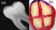
Objectives: In-vitro studies were performed on dentin of extracted human molars to investigate the effectiveness of 9.3 μm CO2 laser irradiation to occlude dentinal tubules. The observed occlusion of dentinal tubules with the irradiation was compared with application of three reagents: 2% Sodium Fluoride gel, an aqueous solution of hydroxyapatite nanoparticles and an equal mix of the two. We show that 9.3 μm CO2 laser irradiation occludes dentinal tubules, and the use of laser irradiation produces better occlusion of the opened tubules compared to the use of topical reagents.
Methods: Nine extracted and cleaned human molars were cut to obtain dentin disks of thickness of 3-5 mm. Each disc was divided into four quarters, and each quarter served as two samples corresponding to irradiated and non-irradiated group counterparts. Five disks were used to study the effect of various laser irradiation energies on the dentinal tubules to find a good pulse fluence for occlusion of the dentinal tubules, and four disks were used for studying the effects of reagents and irradiation at the pulse fluences found in the first part of the study. The samples were irradiated with a beam diameter of 1 mm (1/e2) at 15 Hz pulse repetition rate, scanned automatically using a set of scanning mirrors. Samples were imaged using Scanning Electron Microscope (SEM) which were processed to determine tubule diameter. Safety of the irradiation treatment was investigated on 6 samples by measuring pulpal temperature rise. The effect of three topical reagents corresponding to 2% Sodium Fluoride gel (F), Hydroxyapatite nanoparticles (HA) and an equal mix of F and HA (HAF) on dentinal tubule occlusion was evaluated and compared with the laser irradiation.
Results: In all examined cases, laser irradiation at a fluence of 0.81 J/cm2 resulted in a temperature increase less than 3 °C which is safe, and no surface cracking was observed. There is a threshold pulse fluence of 0.27 J/cm2 above which, laser produced surface melting. At a pulse fluence of 0.81 J/cm2 a layer of recast of melted dentin was formed. Under this layer, peritubular dentin melting and occluding of the dentinal tubules was observed. Application of either F or HA or HAF did not produce visible occlusion effect on open tubules after washing and microbrushing with excess distilled water.
Conclusions: 9.3 μm CO2 laser irradiation on extracted human molar dentin at pulse fluence of 0.81 J./cm2 resulted in tubule area reduction by 97% without rising pulpal temperatures to unsafe levels.
期刊介绍:
Lasers in Medical Science (LIMS) has established itself as the leading international journal in the rapidly expanding field of medical and dental applications of lasers and light. It provides a forum for the publication of papers on the technical, experimental, and clinical aspects of the use of medical lasers, including lasers in surgery, endoscopy, angioplasty, hyperthermia of tumors, and photodynamic therapy. In addition to medical laser applications, LIMS presents high-quality manuscripts on a wide range of dental topics, including aesthetic dentistry, endodontics, orthodontics, and prosthodontics.
The journal publishes articles on the medical and dental applications of novel laser technologies, light delivery systems, sensors to monitor laser effects, basic laser-tissue interactions, and the modeling of laser-tissue interactions. Beyond laser applications, LIMS features articles relating to the use of non-laser light-tissue interactions.


 分享
分享
 求助内容:
求助内容: 应助结果提醒方式:
 扫码关注我们
