Oliver Umney, Joanna Leng, Gianluca Canettieri, Natalia A. Riobo-Del Galdo, Hayley Slaney, Philip Quirke, Michelle Peckham, Alistair Curd
{"title":"Annotation and automated segmentation of single-molecule localisation microscopy data","authors":"Oliver Umney, Joanna Leng, Gianluca Canettieri, Natalia A. Riobo-Del Galdo, Hayley Slaney, Philip Quirke, Michelle Peckham, Alistair Curd","doi":"10.1111/jmi.13349","DOIUrl":null,"url":null,"abstract":"<p>Single Molecule Localisation Microscopy (SMLM) is becoming a widely used technique in cell biology. After processing the images, the molecular localisations are typically stored in a table as <i>xy</i> (or <i>xyz</i>) coordinates, with additional information, such as number of photons, etc. This set of coordinates can be used to generate an image to visualise the molecular distribution, for example, a 2D or 3D histogram of localisations. Many different methods have been devised to analyse SMLM data, among which cluster analysis of the localisations is popular. However, it can be useful to first segment the data, to extract the localisations in a specific region of a cell or in individual cells, prior to downstream analysis. Here we describe a pipeline for annotating localisations in an SMLM dataset in which we compared membrane segmentation approaches, including Otsu thresholding and machine learning models, and subsequent cell segmentation. We used an SMLM dataset derived from dSTORM images of sectioned cell pellets, stained for the membrane proteins EGFR (epidermal growth factor receptor) and EREG (epiregulin) as a test dataset. We found that a Cellpose model retrained on our data performed the best in the membrane segmentation task, allowing us to perform downstream cluster analysis of membrane versus cell interior localisations. We anticipate this will be generally useful for SMLM analysis.</p>","PeriodicalId":16484,"journal":{"name":"Journal of microscopy","volume":"296 3","pages":"214-226"},"PeriodicalIF":1.5000,"publicationDate":"2024-08-02","publicationTypes":"Journal Article","fieldsOfStudy":null,"isOpenAccess":false,"openAccessPdf":"https://onlinelibrary.wiley.com/doi/epdf/10.1111/jmi.13349","citationCount":"0","resultStr":null,"platform":"Semanticscholar","paperid":null,"PeriodicalName":"Journal of microscopy","FirstCategoryId":"5","ListUrlMain":"https://onlinelibrary.wiley.com/doi/10.1111/jmi.13349","RegionNum":4,"RegionCategory":"工程技术","ArticlePicture":[],"TitleCN":null,"AbstractTextCN":null,"PMCID":null,"EPubDate":"","PubModel":"","JCR":"Q3","JCRName":"MICROSCOPY","Score":null,"Total":0}
引用次数: 0
Abstract
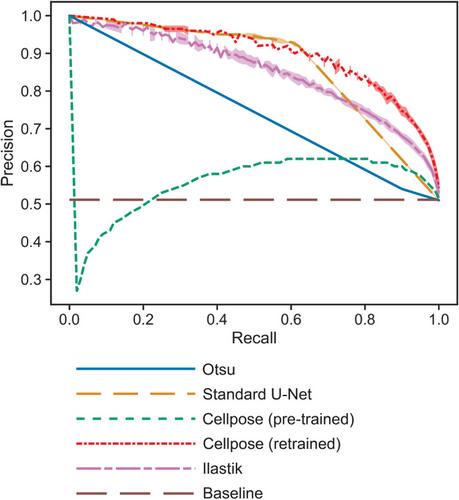
Single Molecule Localisation Microscopy (SMLM) is becoming a widely used technique in cell biology. After processing the images, the molecular localisations are typically stored in a table as xy (or xyz) coordinates, with additional information, such as number of photons, etc. This set of coordinates can be used to generate an image to visualise the molecular distribution, for example, a 2D or 3D histogram of localisations. Many different methods have been devised to analyse SMLM data, among which cluster analysis of the localisations is popular. However, it can be useful to first segment the data, to extract the localisations in a specific region of a cell or in individual cells, prior to downstream analysis. Here we describe a pipeline for annotating localisations in an SMLM dataset in which we compared membrane segmentation approaches, including Otsu thresholding and machine learning models, and subsequent cell segmentation. We used an SMLM dataset derived from dSTORM images of sectioned cell pellets, stained for the membrane proteins EGFR (epidermal growth factor receptor) and EREG (epiregulin) as a test dataset. We found that a Cellpose model retrained on our data performed the best in the membrane segmentation task, allowing us to perform downstream cluster analysis of membrane versus cell interior localisations. We anticipate this will be generally useful for SMLM analysis.
期刊介绍:
The Journal of Microscopy is the oldest journal dedicated to the science of microscopy and the only peer-reviewed publication of the Royal Microscopical Society. It publishes papers that report on the very latest developments in microscopy such as advances in microscopy techniques or novel areas of application. The Journal does not seek to publish routine applications of microscopy or specimen preparation even though the submission may otherwise have a high scientific merit.
The scope covers research in the physical and biological sciences and covers imaging methods using light, electrons, X-rays and other radiations as well as atomic force and near field techniques. Interdisciplinary research is welcome. Papers pertaining to microscopy are also welcomed on optical theory, spectroscopy, novel specimen preparation and manipulation methods and image recording, processing and analysis including dynamic analysis of living specimens.
Publication types include full papers, hot topic fast tracked communications and review articles. Authors considering submitting a review article should contact the editorial office first.


 分享
分享
 求助内容:
求助内容: 应助结果提醒方式:
 扫码关注我们
