Neil MacBeth, Nikos Mardas, Graham Davis, Nikos Donos
{"title":"Healing patterns of alveolar bone following ridge preservation procedures","authors":"Neil MacBeth, Nikos Mardas, Graham Davis, Nikos Donos","doi":"10.1111/clr.14332","DOIUrl":null,"url":null,"abstract":"<div>\n \n \n <section>\n \n <h3> Objectives</h3>\n \n <p>Examine the histomorphometric bone composition, following alveolar ridge preservation techniques and unassisted socket healing.</p>\n </section>\n \n <section>\n \n <h3> Materials and Methods</h3>\n \n <p>Forty-two patients (42) requiring a single rooted tooth extraction were randomly allocated into three groups (<i>n</i> = 14 per group): Group 1: Guided Bone Regeneration (GBR) using deproteinised bovine bone mineral (DBBM) and a porcine collagen membrane; Group 2: Socket Seal (SS) technique using DBBM and a porcine collagen matrix; Group 3: Unassisted socket healing (Control). Trephined bone biopsies were harvested following a 4-month healing period. Forty-two samples underwent Back-Scattered Electrons -Scanning Electron Microscopy (BSE-SEM) imaging, with 15 samples examined using Xray Micro-Tomography (XMT) (<i>n</i> = 6 for each GBR/SS and <i>n</i> = 3 Control). Images were analysed to determine the percentage (%) of connective tissue, new bone formation, residual DBBM particles and direct bone to DBBM particle contact (osseointegration).</p>\n </section>\n \n <section>\n \n <h3> Results</h3>\n \n <p>BSE-SEM analysis demonstrated that new bone formation was higher in the Control (45.89% ± 11.48) compared to both GBR (22.12% ± 12.7/<i>p</i> < .004) and SS (27.62% ± 17.76/<i>p</i> < .005) groups. The connective tissue percentage in GBR (49.72% ± 9), SS (47.81% ± 12.57) and Control (47.81% ± 12.57) groups was similar. GBR (28.17% ± 16.64) and SS (24.37% ± 18.61) groups had similar levels of residual DBBM particles. XMT volumetric analysis indicated a lower level of bone and DBBM particles in all test groups, when matched to the BSE-SEM area measurements. Osseointegration levels (DBBM graft and bone) were recorded at 35.66% (± 9.8) for GBR and 31.18% (± 19.38) for SS.</p>\n </section>\n \n <section>\n \n <h3> Conclusion</h3>\n \n <p>GBR and SS ARP techniques presented with less bone formation when compared to unassisted healing. GBR had more direct contact/osseointegration between the DBBM particles and newly formed bone.</p>\n </section>\n </div>","PeriodicalId":10455,"journal":{"name":"Clinical Oral Implants Research","volume":"35 11","pages":"1452-1466"},"PeriodicalIF":5.3000,"publicationDate":"2024-08-06","publicationTypes":"Journal Article","fieldsOfStudy":null,"isOpenAccess":false,"openAccessPdf":"https://onlinelibrary.wiley.com/doi/epdf/10.1111/clr.14332","citationCount":"0","resultStr":null,"platform":"Semanticscholar","paperid":null,"PeriodicalName":"Clinical Oral Implants Research","FirstCategoryId":"5","ListUrlMain":"https://onlinelibrary.wiley.com/doi/10.1111/clr.14332","RegionNum":1,"RegionCategory":"医学","ArticlePicture":[],"TitleCN":null,"AbstractTextCN":null,"PMCID":null,"EPubDate":"","PubModel":"","JCR":"Q1","JCRName":"DENTISTRY, ORAL SURGERY & MEDICINE","Score":null,"Total":0}
引用次数: 0
Abstract
Objectives
Examine the histomorphometric bone composition, following alveolar ridge preservation techniques and unassisted socket healing.
Materials and Methods
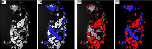
Forty-two patients (42) requiring a single rooted tooth extraction were randomly allocated into three groups (n = 14 per group): Group 1: Guided Bone Regeneration (GBR) using deproteinised bovine bone mineral (DBBM) and a porcine collagen membrane; Group 2: Socket Seal (SS) technique using DBBM and a porcine collagen matrix; Group 3: Unassisted socket healing (Control). Trephined bone biopsies were harvested following a 4-month healing period. Forty-two samples underwent Back-Scattered Electrons -Scanning Electron Microscopy (BSE-SEM) imaging, with 15 samples examined using Xray Micro-Tomography (XMT) (n = 6 for each GBR/SS and n = 3 Control). Images were analysed to determine the percentage (%) of connective tissue, new bone formation, residual DBBM particles and direct bone to DBBM particle contact (osseointegration).
Results
BSE-SEM analysis demonstrated that new bone formation was higher in the Control (45.89% ± 11.48) compared to both GBR (22.12% ± 12.7/p < .004) and SS (27.62% ± 17.76/p < .005) groups. The connective tissue percentage in GBR (49.72% ± 9), SS (47.81% ± 12.57) and Control (47.81% ± 12.57) groups was similar. GBR (28.17% ± 16.64) and SS (24.37% ± 18.61) groups had similar levels of residual DBBM particles. XMT volumetric analysis indicated a lower level of bone and DBBM particles in all test groups, when matched to the BSE-SEM area measurements. Osseointegration levels (DBBM graft and bone) were recorded at 35.66% (± 9.8) for GBR and 31.18% (± 19.38) for SS.
Conclusion
GBR and SS ARP techniques presented with less bone formation when compared to unassisted healing. GBR had more direct contact/osseointegration between the DBBM particles and newly formed bone.
期刊介绍:
Clinical Oral Implants Research conveys scientific progress in the field of implant dentistry and its related areas to clinicians, teachers and researchers concerned with the application of this information for the benefit of patients in need of oral implants. The journal addresses itself to clinicians, general practitioners, periodontists, oral and maxillofacial surgeons and prosthodontists, as well as to teachers, academicians and scholars involved in the education of professionals and in the scientific promotion of the field of implant dentistry.


 分享
分享
 求助内容:
求助内容: 应助结果提醒方式:
 扫码关注我们
