{"title":"Intraoperative margin assessment during radical prostatectomy: is microscopy frozen in time or ready for digital defrost?","authors":"Eoin Dinneen, Ricardo Almeida-Magana, Tarek Al-Hammouri, Iona Fernandes, Nikhil Mayor, Larissa Mendes, Mathias Winkler, Anna Silvanto, Aiman Haider, Alex Freeman, Greg Shaw","doi":"10.1111/his.15290","DOIUrl":null,"url":null,"abstract":"<p>Intraoperative frozen section (IFS) is used with the intention to improve functional and oncological outcomes for patients undergoing radical prostatectomy (RP). High resource requirements of IFS techniques such as NeuroSAFE may preclude widespread adoption, even if there are benefits to patients. Recent advances in fresh-tissue microscopic digital imaging technologies may offer an attractive alternative, and there is a growing body of evidence regarding these technologies. In this narrative review, we discuss some of the familiar limitations of IFS and compare these to the attractive counterpoints of modern digital imaging technologies such as the speed and ease of image generation, the locality of equipment within (or near) the operating room, the ability to maintain tissue integrity, and digital transfer of images. Confocal laser microscopy (CLM) is the modality most frequently reported in the literature for margin assessment during RP. We discuss several imitations and obstacles to widespread dissemination of digital imaging technologies. Among these, we consider how the ‘<i>en-face</i>’ margin perspective will challenge urologists and pathologists to understand afresh the meaning of positive margin significance. As a part of this, discussions on how to describe, categorize, react to, and evaluate these technologies are needed to improve patient outcomes. Limitations of this review include its narrative structure and that the evidence base in this field is relatively immature but developing at pace.</p>","PeriodicalId":13219,"journal":{"name":"Histopathology","volume":"85 5","pages":"716-726"},"PeriodicalIF":4.1000,"publicationDate":"2024-08-05","publicationTypes":"Journal Article","fieldsOfStudy":null,"isOpenAccess":false,"openAccessPdf":"https://onlinelibrary.wiley.com/doi/epdf/10.1111/his.15290","citationCount":"0","resultStr":null,"platform":"Semanticscholar","paperid":null,"PeriodicalName":"Histopathology","FirstCategoryId":"3","ListUrlMain":"https://onlinelibrary.wiley.com/doi/10.1111/his.15290","RegionNum":2,"RegionCategory":"医学","ArticlePicture":[],"TitleCN":null,"AbstractTextCN":null,"PMCID":null,"EPubDate":"","PubModel":"","JCR":"Q2","JCRName":"CELL BIOLOGY","Score":null,"Total":0}
引用次数: 0
Abstract
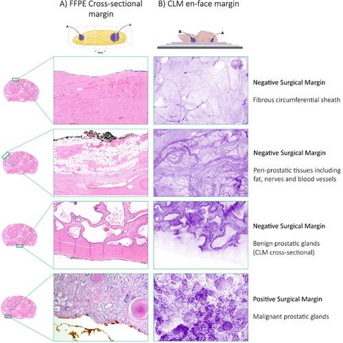
Intraoperative frozen section (IFS) is used with the intention to improve functional and oncological outcomes for patients undergoing radical prostatectomy (RP). High resource requirements of IFS techniques such as NeuroSAFE may preclude widespread adoption, even if there are benefits to patients. Recent advances in fresh-tissue microscopic digital imaging technologies may offer an attractive alternative, and there is a growing body of evidence regarding these technologies. In this narrative review, we discuss some of the familiar limitations of IFS and compare these to the attractive counterpoints of modern digital imaging technologies such as the speed and ease of image generation, the locality of equipment within (or near) the operating room, the ability to maintain tissue integrity, and digital transfer of images. Confocal laser microscopy (CLM) is the modality most frequently reported in the literature for margin assessment during RP. We discuss several imitations and obstacles to widespread dissemination of digital imaging technologies. Among these, we consider how the ‘en-face’ margin perspective will challenge urologists and pathologists to understand afresh the meaning of positive margin significance. As a part of this, discussions on how to describe, categorize, react to, and evaluate these technologies are needed to improve patient outcomes. Limitations of this review include its narrative structure and that the evidence base in this field is relatively immature but developing at pace.
期刊介绍:
Histopathology is an international journal intended to be of practical value to surgical and diagnostic histopathologists, and to investigators of human disease who employ histopathological methods. Our primary purpose is to publish advances in pathology, in particular those applicable to clinical practice and contributing to the better understanding of human disease.


 分享
分享
 求助内容:
求助内容: 应助结果提醒方式:
 扫码关注我们
