{"title":"The effect of laser therapy for the treatment of dentin hypersensitivity on surface roughness and bacterial adhesion.","authors":"Ozge Parlar Oz, İrem Karagozoglu, Ipek Kocer, Nermin Demırkol, Yasemin Zer","doi":"10.1007/s10103-024-04166-0","DOIUrl":null,"url":null,"abstract":"<p><p>The aim of the study was to measure the degree of dentine surface roughness caused by five distinct lasers used to treat dentine hypersensitivity, as well as to evaluate the subsequent bacterial colonization on these irradiated surfaces. Sixty human maxillary premolar teeth without caries or restoration which were extracted for periodontal reasons were used in this study. Five different types of lasers were applied to the root dentin surface. Tested samples were divided into six groups of 10 samples each; control, diode (810 nm), diode (980 nm), Nd: YAG, Er: YAG, and Er, Cr: YSGG laser groups. The arithmetic mean of the surface roughness values (Ra) and the average roughness over a measurement area (Sa) were measured pre- and post-application using any of the laser types. Swab samples were then collected from the dentin surface. Following a 24-hour incubation period at 37 °C, the colony forming units were counted using a stereoscope. The results demonstrated a statistically significant difference in the surface roughness values pre- and post-application (Ra and Sa, respectively) in the Er, Cr: YSGG laser group (p = 0.037,p = 0.007). No significant difference was observed in the other groups (p > 0.05). There was no statistically significant difference in the number of bacterial colonies observed between the test and control groups. Diode and Nd: YAG lasers showed either a decrease or no change in surface roughness; however, the hard tissue lasers (Er: YAG, Er, Cr: YSGG) showed an increase. The Er: YAG and Nd: YAG laser groups exhibited decreased bacterial adhesion compared to the other groups.</p>","PeriodicalId":17978,"journal":{"name":"Lasers in Medical Science","volume":"39 1","pages":"212"},"PeriodicalIF":2.4000,"publicationDate":"2024-08-09","publicationTypes":"Journal Article","fieldsOfStudy":null,"isOpenAccess":false,"openAccessPdf":"https://www.ncbi.nlm.nih.gov/pmc/articles/PMC11315743/pdf/","citationCount":"0","resultStr":null,"platform":"Semanticscholar","paperid":null,"PeriodicalName":"Lasers in Medical Science","FirstCategoryId":"5","ListUrlMain":"https://doi.org/10.1007/s10103-024-04166-0","RegionNum":4,"RegionCategory":"医学","ArticlePicture":[],"TitleCN":null,"AbstractTextCN":null,"PMCID":null,"EPubDate":"","PubModel":"","JCR":"Q3","JCRName":"ENGINEERING, BIOMEDICAL","Score":null,"Total":0}
引用次数: 0
Abstract
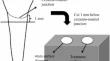
The aim of the study was to measure the degree of dentine surface roughness caused by five distinct lasers used to treat dentine hypersensitivity, as well as to evaluate the subsequent bacterial colonization on these irradiated surfaces. Sixty human maxillary premolar teeth without caries or restoration which were extracted for periodontal reasons were used in this study. Five different types of lasers were applied to the root dentin surface. Tested samples were divided into six groups of 10 samples each; control, diode (810 nm), diode (980 nm), Nd: YAG, Er: YAG, and Er, Cr: YSGG laser groups. The arithmetic mean of the surface roughness values (Ra) and the average roughness over a measurement area (Sa) were measured pre- and post-application using any of the laser types. Swab samples were then collected from the dentin surface. Following a 24-hour incubation period at 37 °C, the colony forming units were counted using a stereoscope. The results demonstrated a statistically significant difference in the surface roughness values pre- and post-application (Ra and Sa, respectively) in the Er, Cr: YSGG laser group (p = 0.037,p = 0.007). No significant difference was observed in the other groups (p > 0.05). There was no statistically significant difference in the number of bacterial colonies observed between the test and control groups. Diode and Nd: YAG lasers showed either a decrease or no change in surface roughness; however, the hard tissue lasers (Er: YAG, Er, Cr: YSGG) showed an increase. The Er: YAG and Nd: YAG laser groups exhibited decreased bacterial adhesion compared to the other groups.
期刊介绍:
Lasers in Medical Science (LIMS) has established itself as the leading international journal in the rapidly expanding field of medical and dental applications of lasers and light. It provides a forum for the publication of papers on the technical, experimental, and clinical aspects of the use of medical lasers, including lasers in surgery, endoscopy, angioplasty, hyperthermia of tumors, and photodynamic therapy. In addition to medical laser applications, LIMS presents high-quality manuscripts on a wide range of dental topics, including aesthetic dentistry, endodontics, orthodontics, and prosthodontics.
The journal publishes articles on the medical and dental applications of novel laser technologies, light delivery systems, sensors to monitor laser effects, basic laser-tissue interactions, and the modeling of laser-tissue interactions. Beyond laser applications, LIMS features articles relating to the use of non-laser light-tissue interactions.


 分享
分享
 求助内容:
求助内容: 应助结果提醒方式:
 扫码关注我们
