George Triantafyllou, Krzysztof Koptas, Nicol Zielinska, Maria Piagkou, Łukasz Olewnik
{"title":"The accessory brachioradialis muscle: prevalence of a rare variant with possible clinical implications.","authors":"George Triantafyllou, Krzysztof Koptas, Nicol Zielinska, Maria Piagkou, Łukasz Olewnik","doi":"10.1007/s00276-024-03462-2","DOIUrl":null,"url":null,"abstract":"<p><strong>Purpose: </strong>The brachioradialis muscle (BRM) belongs to the lateral group of forearm muscles and contributes to the elbow flexion. Accessory brachioradialis muscle (ABRM) or \"brachioradialis accessorius\" represents an uncommon BRM variant, not been enough studied. The present study investigates the prevalence of the ABRM, along with its origin, insertion, and innervation.</p><p><strong>Materials: </strong>Eighty-three upper limbs were meticulously dissected at the arm, forearm, and cubital fossa to investigate the ABRM presence. When the variant muscle was identified, morphometric measurements were obtained.</p><p><strong>Results: </strong>The ABRM was identified in two upper limbs (2/83, 2.4%), in a male cadaver, bilaterally. Its origin was located along with the typical BRM, and its insertion was identified into the anterior surface of the radius (proximal third). The ABRM was innervated by the radial nerve, coursing posteriorly (deeply).</p><p><strong>Conclusions: </strong>In the current study, the variant muscle was observed in 2.4%. Radial nerve compression, at the forearm, is not an uncommon entrapment neuropathy. The relationship between the radial nerve and the ABRM could precipitate radial neuropathy.</p>","PeriodicalId":49461,"journal":{"name":"Surgical and Radiologic Anatomy","volume":" ","pages":"1709-1714"},"PeriodicalIF":1.2000,"publicationDate":"2024-10-01","publicationTypes":"Journal Article","fieldsOfStudy":null,"isOpenAccess":false,"openAccessPdf":"","citationCount":"0","resultStr":null,"platform":"Semanticscholar","paperid":null,"PeriodicalName":"Surgical and Radiologic Anatomy","FirstCategoryId":"3","ListUrlMain":"https://doi.org/10.1007/s00276-024-03462-2","RegionNum":4,"RegionCategory":"医学","ArticlePicture":[],"TitleCN":null,"AbstractTextCN":null,"PMCID":null,"EPubDate":"2024/8/13 0:00:00","PubModel":"Epub","JCR":"Q2","JCRName":"Medicine","Score":null,"Total":0}
引用次数: 0
Abstract
Purpose: The brachioradialis muscle (BRM) belongs to the lateral group of forearm muscles and contributes to the elbow flexion. Accessory brachioradialis muscle (ABRM) or "brachioradialis accessorius" represents an uncommon BRM variant, not been enough studied. The present study investigates the prevalence of the ABRM, along with its origin, insertion, and innervation.
Materials: Eighty-three upper limbs were meticulously dissected at the arm, forearm, and cubital fossa to investigate the ABRM presence. When the variant muscle was identified, morphometric measurements were obtained.
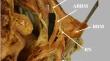
Results: The ABRM was identified in two upper limbs (2/83, 2.4%), in a male cadaver, bilaterally. Its origin was located along with the typical BRM, and its insertion was identified into the anterior surface of the radius (proximal third). The ABRM was innervated by the radial nerve, coursing posteriorly (deeply).
Conclusions: In the current study, the variant muscle was observed in 2.4%. Radial nerve compression, at the forearm, is not an uncommon entrapment neuropathy. The relationship between the radial nerve and the ABRM could precipitate radial neuropathy.
期刊介绍:
Anatomy is a morphological science which cannot fail to interest the clinician. The practical application of anatomical research to clinical problems necessitates special adaptation and selectivity in choosing from numerous international works. Although there is a tendency to believe that meaningful advances in anatomy are unlikely, constant revision is necessary. Surgical and Radiologic Anatomy, the first international journal of Clinical anatomy has been created in this spirit.
Its goal is to serve clinicians, regardless of speciality-physicians, surgeons, radiologists or other specialists-as an indispensable aid with which they can improve their knowledge of anatomy. Each issue includes: Original papers, review articles, articles on the anatomical bases of medical, surgical and radiological techniques, articles of normal radiologic anatomy, brief reviews of anatomical publications of clinical interest.
Particular attention is given to high quality illustrations, which are indispensable for a better understanding of anatomical problems.
Surgical and Radiologic Anatomy is a journal written by anatomists for clinicians with a special interest in anatomy.


 分享
分享
 求助内容:
求助内容: 应助结果提醒方式:
 扫码关注我们
