Eric L Tung, Amine El Kandoussi, Steven J Staffa, Daniel I Rosenthal, Connie Y Chang
{"title":"Elongated morphology of osteoid osteoma is associated with radiofrequency ablation failure in children.","authors":"Eric L Tung, Amine El Kandoussi, Steven J Staffa, Daniel I Rosenthal, Connie Y Chang","doi":"10.1007/s00256-024-04776-3","DOIUrl":null,"url":null,"abstract":"<p><strong>Objective: </strong>To compare the frequency of elongated morphology of osteoid osteoma (OO) in children compared to adolescents and to determine if this elongated morphology is associated with radiofrequency ablation treatment failure.</p><p><strong>Materials and methods: </strong>Retrospective review of first-time CT-guided radiofrequency ablation performed for presumed OO in patients < 21 years old between 1990 and 2023. Children were considered 0 to 10 years old, and adolescents were considered 11 to 20 years old. Treatment failure was considered symptomatic recurrence requiring follow-up intervention. The largest tumor dimensions in three orthogonal planes were measured using multiplanar reformatted technology. Maximum tumor dimension, tumor volume, and eccentricity index were calculated. Elongated morphology criteria were (a) largest dimension > 10 mm and (b) eccentricity index ≥ 3. Lesion locations were recorded. Statistical analyses included the chi-square test, Fisher's exact test, nonparametric Wilcoxon rank-sum test, receiver operating characteristic analysis, and Spearman's nonparametric rank correlation.</p><p><strong>Results: </strong>Of 366 included patients (median 15 years, IQR 11-18 years, 254 male), there were 86 (23.5%) children, 280 (76.5%) adolescents, and 24 (6.6%) cases of treatment failure. Elongated morphology was more common in children (19.7%) than adolescents (8.6%) (p = 0.004) and associated with younger age (p = 0.009). Elongated morphology was associated with treatment failure in children (p = 0.045) but not adolescents (p > .99) or all patients (p = 0.17). Treatment failure was not associated with age, largest dimension, eccentricity index, volume, or location.</p><p><strong>Conclusion: </strong>Elongated morphology of OO is associated with younger age and radiofrequency ablation treatment failure in children. Identifying this morphology may assist with counseling and treatment planning.</p>","PeriodicalId":21783,"journal":{"name":"Skeletal Radiology","volume":" ","pages":"553-561"},"PeriodicalIF":2.2000,"publicationDate":"2025-03-01","publicationTypes":"Journal Article","fieldsOfStudy":null,"isOpenAccess":false,"openAccessPdf":"","citationCount":"0","resultStr":null,"platform":"Semanticscholar","paperid":null,"PeriodicalName":"Skeletal Radiology","FirstCategoryId":"3","ListUrlMain":"https://doi.org/10.1007/s00256-024-04776-3","RegionNum":3,"RegionCategory":"医学","ArticlePicture":[],"TitleCN":null,"AbstractTextCN":null,"PMCID":null,"EPubDate":"2024/8/24 0:00:00","PubModel":"Epub","JCR":"Q2","JCRName":"ORTHOPEDICS","Score":null,"Total":0}
引用次数: 0
Abstract
Objective: To compare the frequency of elongated morphology of osteoid osteoma (OO) in children compared to adolescents and to determine if this elongated morphology is associated with radiofrequency ablation treatment failure.
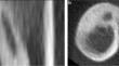
Materials and methods: Retrospective review of first-time CT-guided radiofrequency ablation performed for presumed OO in patients < 21 years old between 1990 and 2023. Children were considered 0 to 10 years old, and adolescents were considered 11 to 20 years old. Treatment failure was considered symptomatic recurrence requiring follow-up intervention. The largest tumor dimensions in three orthogonal planes were measured using multiplanar reformatted technology. Maximum tumor dimension, tumor volume, and eccentricity index were calculated. Elongated morphology criteria were (a) largest dimension > 10 mm and (b) eccentricity index ≥ 3. Lesion locations were recorded. Statistical analyses included the chi-square test, Fisher's exact test, nonparametric Wilcoxon rank-sum test, receiver operating characteristic analysis, and Spearman's nonparametric rank correlation.
Results: Of 366 included patients (median 15 years, IQR 11-18 years, 254 male), there were 86 (23.5%) children, 280 (76.5%) adolescents, and 24 (6.6%) cases of treatment failure. Elongated morphology was more common in children (19.7%) than adolescents (8.6%) (p = 0.004) and associated with younger age (p = 0.009). Elongated morphology was associated with treatment failure in children (p = 0.045) but not adolescents (p > .99) or all patients (p = 0.17). Treatment failure was not associated with age, largest dimension, eccentricity index, volume, or location.
Conclusion: Elongated morphology of OO is associated with younger age and radiofrequency ablation treatment failure in children. Identifying this morphology may assist with counseling and treatment planning.
期刊介绍:
Skeletal Radiology provides a forum for the dissemination of current knowledge and information dealing with disorders of the musculoskeletal system including the spine. While emphasizing the radiological aspects of the many varied skeletal abnormalities, the journal also adopts an interdisciplinary approach, reflecting the membership of the International Skeletal Society. Thus, the anatomical, pathological, physiological, clinical, metabolic and epidemiological aspects of the many entities affecting the skeleton receive appropriate consideration.
This is the Journal of the International Skeletal Society and the Official Journal of the Society of Skeletal Radiology and the Australasian Musculoskelelal Imaging Group.


 分享
分享
 求助内容:
求助内容: 应助结果提醒方式:
 扫码关注我们
