To discuss the usefulness of ultrasonography (US) in the diagnosis and management of pediatric head and neck lymphatic malformations (HNLMs).
We conducted a retrospective analysis of 140 children who were referred to our hospital for the treatment of HNLMs.
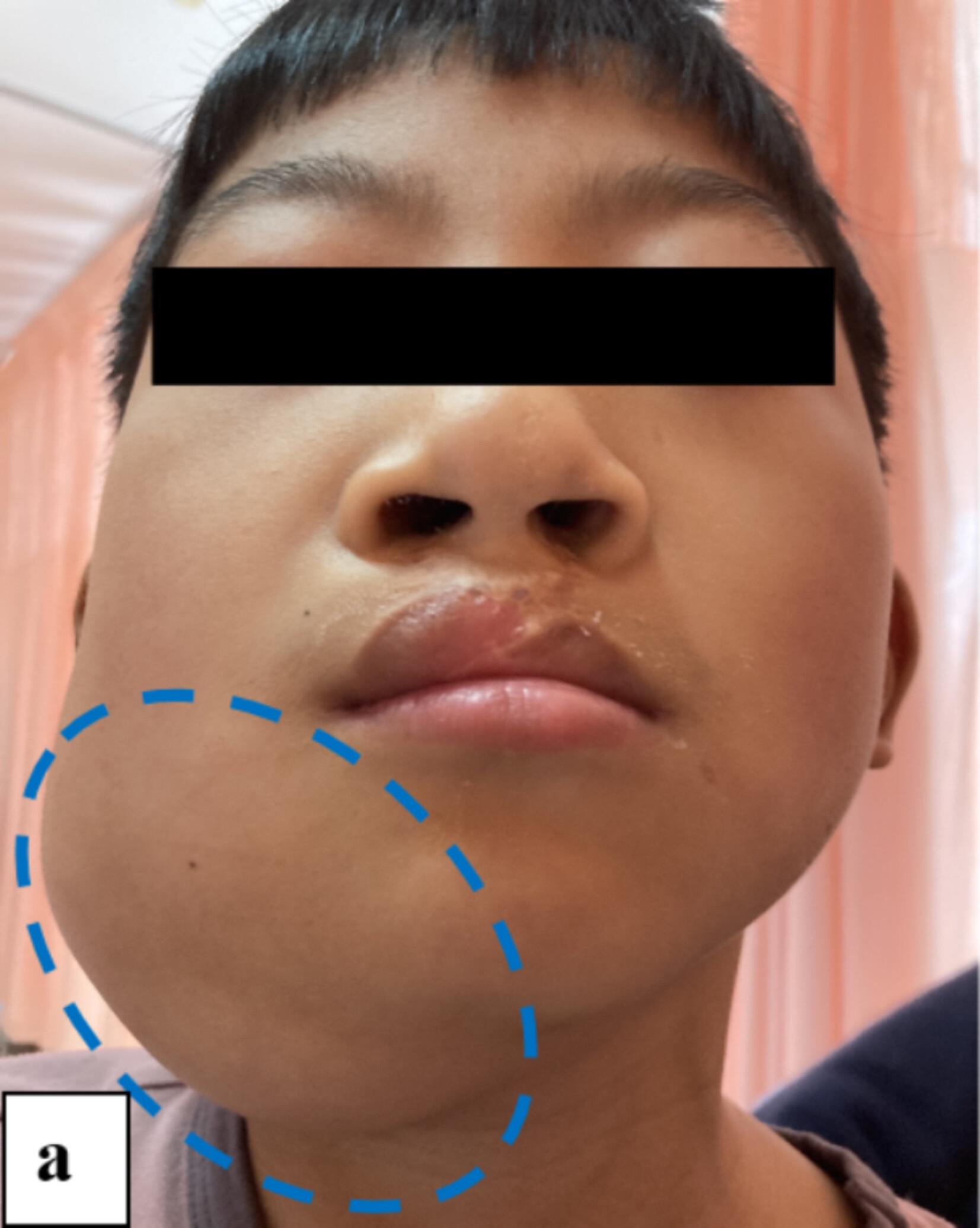
The median age at presentation was 12 months (1 day–171 months; 66.4% under 2 years old; 35.7% neonatus). The majority clinical presentations were asymptomatic mass (65.7%, 92/140) and cosmetic deformity (25.7%, 36/140). HNLMs involved the neck accounting for 65.7% (92/140), especially posterior cervical trigone (22.1%, 31/140), and submandibular (20.0%, 28/140). The US diagnostic accuracy was 91.4% (128/140). Their boundary with the surrounding tissues was usually clear (87.9%, 123/140), whereas the shape was mostly irregular (97.1%, 136/140). Based on surgical findings, there were 67 pure HNLMs and 73 intracystic hemorrhage. Between the two groups, there were statistical differences in capsule contents (χ2 = 7.8299, p = 0.0051), flocculent echo floating (χ2 = 21.2964, p < 0.0001), overlying skin (χ2 = 9.0498, p = 0.0026), and palpation (χ2 = 13.4058, p = 0.0003).
US typically reveals the lesion with clear boundary, irregular morphology, anechoic contents, no blood flow signal, and echoic intracapsular septum with blood flow signal. In contrast, bluish appearance, tensional palpation, and capsule contents with low/mixed echo or flocculent echo floating may indicate intracystic hemorrhage.


 分享
分享
 求助内容:
求助内容: 应助结果提醒方式:
 扫码关注我们
