{"title":"Myocardial calcification: case reports and a systematic review.","authors":"Takashi Kido, Kazuki Tanimoto, Takuji Watanabe, Masaki Taira, Jun Narita, Hidekazu Ishida, Ryo Ishii, Takayoshi Ueno, Shigeru Miyagawa","doi":"10.1093/ehjimp/qyae079","DOIUrl":null,"url":null,"abstract":"<p><strong>Aims: </strong>Myocardial calcification is an unusual condition in which excess calcium is deposited in the myocardium. Herein, we report two cases of myocardial calcification from our clinical experience. Furthermore, we conduct a systematic review to examine the clinical course and associated pathologies of myocardial calcification.</p><p><strong>Methods and results: </strong>This systematic review was registered in PROSPERO (CRD42023463285). PubMed and Scopus were searched according to the following inclusion criteria: (i) case reports or case series describing patients with myocardial calcification; (ii) diagnosis of myocardial calcification by computed tomography (CT); (iii) adequate description of patients, including their chief complaint, medical history, evaluations, and treatments; and (iv) publication in English. Among the 75 patients, 24 had sepsis, 14 had myocarditis, and 37 had other pathologies. The mortality rate was 33% for patients with sepsis, 14% for patients with myocarditis, and 11% for patients with other pathologies. Follow-up CT findings beyond 2 years were reported in six patients, showing that the CT findings of myocardial calcification persisted but subsided over time. Autopsy was performed in seven patients, and extensive interstitial fibrosis and collection of inflammatory cells were observed in patients with myocarditis, sepsis, and ischaemic heart disease.</p><p><strong>Conclusion: </strong>While various medical conditions can cause myocardial calcification, accompanying conditions commonly reported with myocardial calcification were sepsis and myocarditis. The CT findings of myocardial calcification tend to regress over time if the underlying disease can be treated.</p>","PeriodicalId":94317,"journal":{"name":"European heart journal. Imaging methods and practice","volume":"2 3","pages":"qyae079"},"PeriodicalIF":0.0000,"publicationDate":"2024-07-30","publicationTypes":"Journal Article","fieldsOfStudy":null,"isOpenAccess":false,"openAccessPdf":"https://www.ncbi.nlm.nih.gov/pmc/articles/PMC11367960/pdf/","citationCount":"0","resultStr":null,"platform":"Semanticscholar","paperid":null,"PeriodicalName":"European heart journal. Imaging methods and practice","FirstCategoryId":"1085","ListUrlMain":"https://doi.org/10.1093/ehjimp/qyae079","RegionNum":0,"RegionCategory":null,"ArticlePicture":[],"TitleCN":null,"AbstractTextCN":null,"PMCID":null,"EPubDate":"2024/7/1 0:00:00","PubModel":"eCollection","JCR":"","JCRName":"","Score":null,"Total":0}
引用次数: 0
Abstract
Aims: Myocardial calcification is an unusual condition in which excess calcium is deposited in the myocardium. Herein, we report two cases of myocardial calcification from our clinical experience. Furthermore, we conduct a systematic review to examine the clinical course and associated pathologies of myocardial calcification.
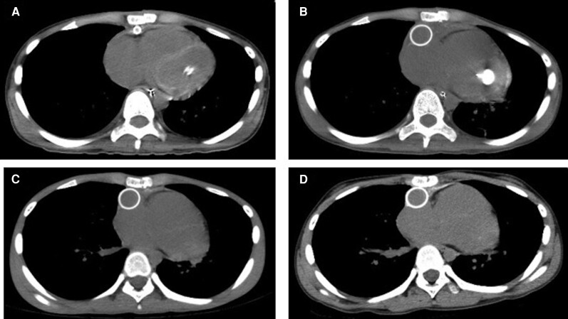
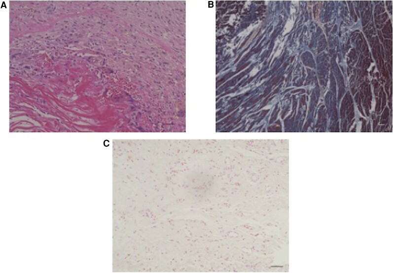
Methods and results: This systematic review was registered in PROSPERO (CRD42023463285). PubMed and Scopus were searched according to the following inclusion criteria: (i) case reports or case series describing patients with myocardial calcification; (ii) diagnosis of myocardial calcification by computed tomography (CT); (iii) adequate description of patients, including their chief complaint, medical history, evaluations, and treatments; and (iv) publication in English. Among the 75 patients, 24 had sepsis, 14 had myocarditis, and 37 had other pathologies. The mortality rate was 33% for patients with sepsis, 14% for patients with myocarditis, and 11% for patients with other pathologies. Follow-up CT findings beyond 2 years were reported in six patients, showing that the CT findings of myocardial calcification persisted but subsided over time. Autopsy was performed in seven patients, and extensive interstitial fibrosis and collection of inflammatory cells were observed in patients with myocarditis, sepsis, and ischaemic heart disease.
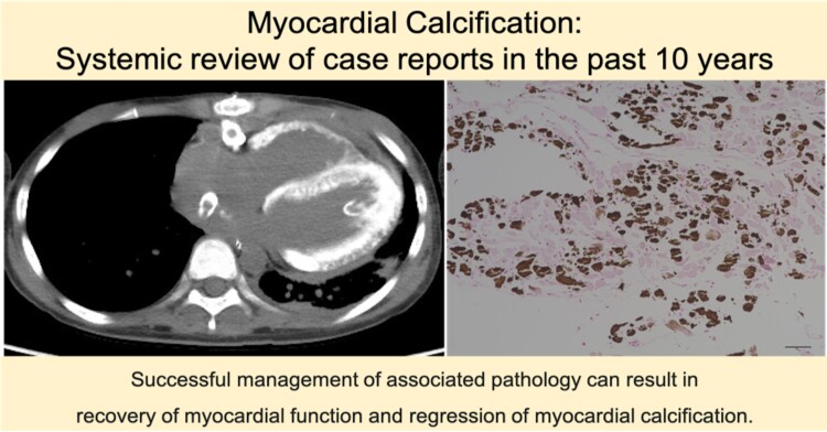
Conclusion: While various medical conditions can cause myocardial calcification, accompanying conditions commonly reported with myocardial calcification were sepsis and myocarditis. The CT findings of myocardial calcification tend to regress over time if the underlying disease can be treated.


 分享
分享

 求助内容:
求助内容: 应助结果提醒方式:
 扫码关注我们
