The apparent diffusion coefficient based on small-field DWI is superior to T2-weighted imaging in evaluating neurological dysfunction of degenerative cervical myelopathy.
Xiao-Nan Tian, Sheng-Nan Li, Bao-Gen Zhao, Ning Wang, Ting Gao, Li Zhang
{"title":"The apparent diffusion coefficient based on small-field DWI is superior to T2-weighted imaging in evaluating neurological dysfunction of degenerative cervical myelopathy.","authors":"Xiao-Nan Tian, Sheng-Nan Li, Bao-Gen Zhao, Ning Wang, Ting Gao, Li Zhang","doi":"10.1007/s00586-024-08411-6","DOIUrl":null,"url":null,"abstract":"<p><strong>Purpose: </strong>To investigate the clinical application of zonally magnified oblique multislice (ZOOM) imaging technology in patients with degenerative cervical myelopathy (DCM) and compare it with T2WI imaging.</p><p><strong>Methods: </strong>A total of 111 patients diagnosed with DCM were recruited. According to mJOA, patients with DCM were divided into ND + group with neurological dysfunction and ND- group without neurological dysfunction. Routine MRI and ZOOM-DWI were performed on 3.0 T MRI to obtain sagittal T2WI and apparent diffusion coefficient (ADC) diagram. ADC values of the narrow segment and its adjacent upper and lower segments were measured, and compared between the ND + and ND- groups. The correlation between ADC value of cervical spinal cord and mJOA score was analyzed. Additionally, ROC curves were plotted to calculate the AUC values.</p><p><strong>Results: </strong>The comparison between ND + and ND- groups shows that there are significant differences in mJOA score, T2WI, anteroposterior diameter of spinal canal, ADC values of narrow, upper and lower segment (P < 0.05). In ND + group, there is a significant difference between ADC values of the narrow and its upper and lower segments (P < 0.001), while with no significant difference in ADC values of the upper and lower segments (P > 0.05). Results of correlation analysis indicate that in the ND + group, neurological dysfunction evaluated by mJOA scores is correlated with increased ADC values of the narrow segment (r = -0.52, P < 0.001), but not significantly correlated with ADC values of the upper and lower segments. Furthermore, T2WI, anteroposterior diameter of the spinal canal, and cervical cord ADC values all has diagnostic efficacy in evaluating neurological dysfunction in DCM (AUC > 0.5, P < 0.05), with the ADC value of the narrow segment being optimal.</p><p><strong>Conclusion: </strong>The ADC value of spinal cord obtained by small-field ZOOM-DWI can be used to evaluate neurological dysfunction in DCM, and is superior to traditional T2WI.</p>","PeriodicalId":12323,"journal":{"name":"European Spine Journal","volume":" ","pages":"3949-3956"},"PeriodicalIF":2.7000,"publicationDate":"2024-10-01","publicationTypes":"Journal Article","fieldsOfStudy":null,"isOpenAccess":false,"openAccessPdf":"","citationCount":"0","resultStr":null,"platform":"Semanticscholar","paperid":null,"PeriodicalName":"European Spine Journal","FirstCategoryId":"3","ListUrlMain":"https://doi.org/10.1007/s00586-024-08411-6","RegionNum":3,"RegionCategory":"医学","ArticlePicture":[],"TitleCN":null,"AbstractTextCN":null,"PMCID":null,"EPubDate":"2024/9/4 0:00:00","PubModel":"Epub","JCR":"Q2","JCRName":"CLINICAL NEUROLOGY","Score":null,"Total":0}
引用次数: 0
Abstract
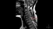
Purpose: To investigate the clinical application of zonally magnified oblique multislice (ZOOM) imaging technology in patients with degenerative cervical myelopathy (DCM) and compare it with T2WI imaging.
Methods: A total of 111 patients diagnosed with DCM were recruited. According to mJOA, patients with DCM were divided into ND + group with neurological dysfunction and ND- group without neurological dysfunction. Routine MRI and ZOOM-DWI were performed on 3.0 T MRI to obtain sagittal T2WI and apparent diffusion coefficient (ADC) diagram. ADC values of the narrow segment and its adjacent upper and lower segments were measured, and compared between the ND + and ND- groups. The correlation between ADC value of cervical spinal cord and mJOA score was analyzed. Additionally, ROC curves were plotted to calculate the AUC values.
Results: The comparison between ND + and ND- groups shows that there are significant differences in mJOA score, T2WI, anteroposterior diameter of spinal canal, ADC values of narrow, upper and lower segment (P < 0.05). In ND + group, there is a significant difference between ADC values of the narrow and its upper and lower segments (P < 0.001), while with no significant difference in ADC values of the upper and lower segments (P > 0.05). Results of correlation analysis indicate that in the ND + group, neurological dysfunction evaluated by mJOA scores is correlated with increased ADC values of the narrow segment (r = -0.52, P < 0.001), but not significantly correlated with ADC values of the upper and lower segments. Furthermore, T2WI, anteroposterior diameter of the spinal canal, and cervical cord ADC values all has diagnostic efficacy in evaluating neurological dysfunction in DCM (AUC > 0.5, P < 0.05), with the ADC value of the narrow segment being optimal.
Conclusion: The ADC value of spinal cord obtained by small-field ZOOM-DWI can be used to evaluate neurological dysfunction in DCM, and is superior to traditional T2WI.
期刊介绍:
"European Spine Journal" is a publication founded in response to the increasing trend toward specialization in spinal surgery and spinal pathology in general. The Journal is devoted to all spine related disciplines, including functional and surgical anatomy of the spine, biomechanics and pathophysiology, diagnostic procedures, and neurology, surgery and outcomes. The aim of "European Spine Journal" is to support the further development of highly innovative spine treatments including but not restricted to surgery and to provide an integrated and balanced view of diagnostic, research and treatment procedures as well as outcomes that will enhance effective collaboration among specialists worldwide. The “European Spine Journal” also participates in education by means of videos, interactive meetings and the endorsement of educative efforts.
Official publication of EUROSPINE, The Spine Society of Europe


 分享
分享
 求助内容:
求助内容: 应助结果提醒方式:
 扫码关注我们
