Yang Li, Xian Shao, Li-Juan Dai, Meng Yu, Meng-Di Cong, Jun-Yi Sun, Shuo Pan, Gao-Feng Shi, An-Du Zhang, Hui Liu
{"title":"Development of a prognostic nomogram for esophageal squamous cell carcinoma patients received radiotherapy based on clinical risk factors.","authors":"Yang Li, Xian Shao, Li-Juan Dai, Meng Yu, Meng-Di Cong, Jun-Yi Sun, Shuo Pan, Gao-Feng Shi, An-Du Zhang, Hui Liu","doi":"10.3389/fonc.2024.1429790","DOIUrl":null,"url":null,"abstract":"<p><strong>Purpose: </strong>The goal of the study was to create a nomogram based on clinical risk factors to forecast the rate of locoregional recurrence-free survival (LRFS) in patients with esophageal squamous cell carcinoma (ESCC) who underwent radiotherapy (RT).</p><p><strong>Methods: </strong>In this study, 574 ESCC patients were selected as participants. Following radiotherapy, subjects were divided into training and validation groups at a 7:3 ratio. The nomogram was established in the training group using Cox regression. Performance validation was conducted in the validation group, assessing predictability through the C-index and AUC curve, calibration via the Hosmer-Lemeshow (H-L) test, and evaluating clinical applicability using decision curve analysis (DCA).</p><p><strong>Results: </strong>T stage, N stage, gross tumor volume (GTV) dose, location, maximal wall thickness (MWT) after RT, node size (NS) after RT, Δ computer tomography (CT) value, and chemotherapy were found to be independent risk factors that impacted LRFS by multivariate cox analysis, and the findings could be utilized to create a nomogram and forecast LRFS. the area under the receiver operating characteristic (AUC) curve and C-index show that for training and validation groups, the prediction result of LRFS using nomogram was more accurate than that of TNM. The LRFS in both groups was consistent with the nomogram according to the H-L test. The DCA curve demonstrated that the nomogram had a good prediction effect both in the groups for training and validation. The nomogram was used to assign ESCC patients to three risk levels: low, medium, or high. There were substantial variations in LRFS between risk categories in both the training and validation groups (p<0.001, p=0.003).</p><p><strong>Conclusions: </strong>For ESCC patients who received radiotherapy, the nomogram based on clinical risk factors could reliably predict the LRFS.</p>","PeriodicalId":12482,"journal":{"name":"Frontiers in Oncology","volume":"14 ","pages":"1429790"},"PeriodicalIF":3.5000,"publicationDate":"2024-08-22","publicationTypes":"Journal Article","fieldsOfStudy":null,"isOpenAccess":false,"openAccessPdf":"https://www.ncbi.nlm.nih.gov/pmc/articles/PMC11374629/pdf/","citationCount":"0","resultStr":null,"platform":"Semanticscholar","paperid":null,"PeriodicalName":"Frontiers in Oncology","FirstCategoryId":"3","ListUrlMain":"https://doi.org/10.3389/fonc.2024.1429790","RegionNum":3,"RegionCategory":"医学","ArticlePicture":[],"TitleCN":null,"AbstractTextCN":null,"PMCID":null,"EPubDate":"2024/1/1 0:00:00","PubModel":"eCollection","JCR":"Q2","JCRName":"ONCOLOGY","Score":null,"Total":0}
引用次数: 0
Abstract
Purpose: The goal of the study was to create a nomogram based on clinical risk factors to forecast the rate of locoregional recurrence-free survival (LRFS) in patients with esophageal squamous cell carcinoma (ESCC) who underwent radiotherapy (RT).
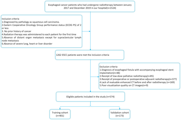
Methods: In this study, 574 ESCC patients were selected as participants. Following radiotherapy, subjects were divided into training and validation groups at a 7:3 ratio. The nomogram was established in the training group using Cox regression. Performance validation was conducted in the validation group, assessing predictability through the C-index and AUC curve, calibration via the Hosmer-Lemeshow (H-L) test, and evaluating clinical applicability using decision curve analysis (DCA).
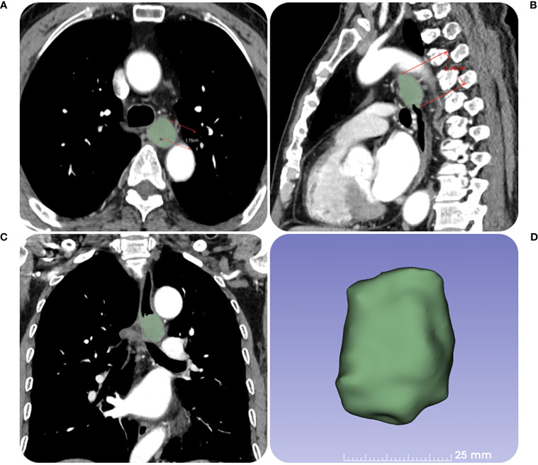
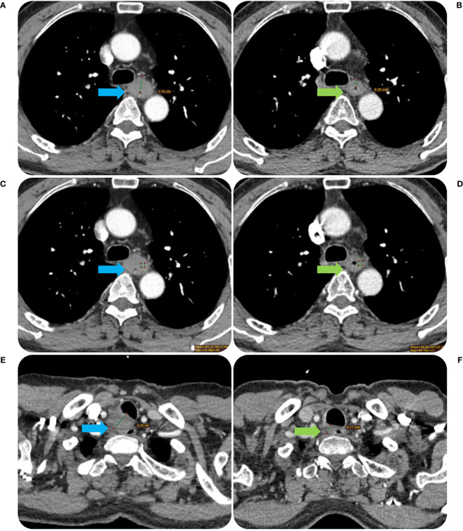
Results: T stage, N stage, gross tumor volume (GTV) dose, location, maximal wall thickness (MWT) after RT, node size (NS) after RT, Δ computer tomography (CT) value, and chemotherapy were found to be independent risk factors that impacted LRFS by multivariate cox analysis, and the findings could be utilized to create a nomogram and forecast LRFS. the area under the receiver operating characteristic (AUC) curve and C-index show that for training and validation groups, the prediction result of LRFS using nomogram was more accurate than that of TNM. The LRFS in both groups was consistent with the nomogram according to the H-L test. The DCA curve demonstrated that the nomogram had a good prediction effect both in the groups for training and validation. The nomogram was used to assign ESCC patients to three risk levels: low, medium, or high. There were substantial variations in LRFS between risk categories in both the training and validation groups (p<0.001, p=0.003).
Conclusions: For ESCC patients who received radiotherapy, the nomogram based on clinical risk factors could reliably predict the LRFS.
期刊介绍:
Cancer Imaging and Diagnosis is dedicated to the publication of results from clinical and research studies applied to cancer diagnosis and treatment. The section aims to publish studies from the entire field of cancer imaging: results from routine use of clinical imaging in both radiology and nuclear medicine, results from clinical trials, experimental molecular imaging in humans and small animals, research on new contrast agents in CT, MRI, ultrasound, publication of new technical applications and processing algorithms to improve the standardization of quantitative imaging and image guided interventions for the diagnosis and treatment of cancer.


 分享
分享
 求助内容:
求助内容: 应助结果提醒方式:
 扫码关注我们
