Eric D. Nguyen, Chien-Kuang Cornelia Ding, Sarah E. Umetsu, Linda D. Ferrell, Kwun Wah Wen
{"title":"Glutamine synthetase staining patterns in cirrhosis","authors":"Eric D. Nguyen, Chien-Kuang Cornelia Ding, Sarah E. Umetsu, Linda D. Ferrell, Kwun Wah Wen","doi":"10.1016/j.humpath.2024.105655","DOIUrl":null,"url":null,"abstract":"<div><p>Advanced liver fibrosis can regress following the elimination of causative injuries. Glutamine synthetase (GS) immunohistochemical expression is normally in centrizonal perivenular hepatocytes but can be present in periportal hepatocytes in cases of regressed cirrhosis. This study identified periportal staining and investigated the spectrum of GS staining patterns seen in a range of cirrhotic livers with varying disease processes. The hematoxylin and eosin and GS-stained slides of 88 liver resection/explant specimens with advanced fibrosis cases by different causes were reviewed, and trichrome and orcein stains were used to classify cases as progressive, indeterminate, or regressive. Periportal GS staining was seen in 97% of regressive cases and 84% progressive or indeterminate cases. Liver resection specimens with periportal GS staining showed a variety of patterns, including predominantly perivenular, predominantly periseptal, and perinodular staining. The GS periseptal pattern is more common in regressed cirrhosis compared to progressive cases. The perinodular staining was seen in 16 cases resulting from various etiologies, including biliary atresia, steatotic liver disease, primary biliary cholangitis, and viral hepatitis, 75% of which demonstrated cholestasis. This study further subclassified GS staining patterns of “periportal” pattern in cirrhotic liver. Compared to orcein/trichrome staining, GS immunohistochemical staining is not as useful in distinguishing regressed cases from non-regressed cases.</p></div>","PeriodicalId":13062,"journal":{"name":"Human pathology","volume":"153 ","pages":"Article 105655"},"PeriodicalIF":2.6000,"publicationDate":"2024-11-01","publicationTypes":"Journal Article","fieldsOfStudy":null,"isOpenAccess":false,"openAccessPdf":"","citationCount":"0","resultStr":null,"platform":"Semanticscholar","paperid":null,"PeriodicalName":"Human pathology","FirstCategoryId":"3","ListUrlMain":"https://www.sciencedirect.com/science/article/pii/S0046817724001643","RegionNum":2,"RegionCategory":"医学","ArticlePicture":[],"TitleCN":null,"AbstractTextCN":null,"PMCID":null,"EPubDate":"2024/9/6 0:00:00","PubModel":"Epub","JCR":"Q2","JCRName":"PATHOLOGY","Score":null,"Total":0}
引用次数: 0
Abstract
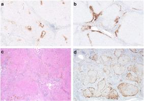
Advanced liver fibrosis can regress following the elimination of causative injuries. Glutamine synthetase (GS) immunohistochemical expression is normally in centrizonal perivenular hepatocytes but can be present in periportal hepatocytes in cases of regressed cirrhosis. This study identified periportal staining and investigated the spectrum of GS staining patterns seen in a range of cirrhotic livers with varying disease processes. The hematoxylin and eosin and GS-stained slides of 88 liver resection/explant specimens with advanced fibrosis cases by different causes were reviewed, and trichrome and orcein stains were used to classify cases as progressive, indeterminate, or regressive. Periportal GS staining was seen in 97% of regressive cases and 84% progressive or indeterminate cases. Liver resection specimens with periportal GS staining showed a variety of patterns, including predominantly perivenular, predominantly periseptal, and perinodular staining. The GS periseptal pattern is more common in regressed cirrhosis compared to progressive cases. The perinodular staining was seen in 16 cases resulting from various etiologies, including biliary atresia, steatotic liver disease, primary biliary cholangitis, and viral hepatitis, 75% of which demonstrated cholestasis. This study further subclassified GS staining patterns of “periportal” pattern in cirrhotic liver. Compared to orcein/trichrome staining, GS immunohistochemical staining is not as useful in distinguishing regressed cases from non-regressed cases.
期刊介绍:
Human Pathology is designed to bring information of clinicopathologic significance to human disease to the laboratory and clinical physician. It presents information drawn from morphologic and clinical laboratory studies with direct relevance to the understanding of human diseases. Papers published concern morphologic and clinicopathologic observations, reviews of diseases, analyses of problems in pathology, significant collections of case material and advances in concepts or techniques of value in the analysis and diagnosis of disease. Theoretical and experimental pathology and molecular biology pertinent to human disease are included. This critical journal is well illustrated with exceptional reproductions of photomicrographs and microscopic anatomy.


 分享
分享
 求助内容:
求助内容: 应助结果提醒方式:
 扫码关注我们
