Periaqueductal gray connectivity in spinal cord injury-induced neuropathic pain

IF 2.3 4区 医学 Q3 CLINICAL NEUROLOGY Journal of Neuroimaging Pub Date : 2024-09-09 DOI:10.1111/jon.13237
Omid Shoraka, Mashaal Syed, Shreya Mandloi, Sara Thalheimer, Sara Naghizadeh Kashani, Joshua E. Heller, Feroze B. Mohamed, Ashwini D. Sharan, Kiran S. Talekar, Caio M. Matias, James S. Harrop, Laura Krisa, Mahdi Alizadeh
{"title":"Periaqueductal gray connectivity in spinal cord injury-induced neuropathic pain","authors":"Omid Shoraka,&nbsp;Mashaal Syed,&nbsp;Shreya Mandloi,&nbsp;Sara Thalheimer,&nbsp;Sara Naghizadeh Kashani,&nbsp;Joshua E. Heller,&nbsp;Feroze B. Mohamed,&nbsp;Ashwini D. Sharan,&nbsp;Kiran S. Talekar,&nbsp;Caio M. Matias,&nbsp;James S. Harrop,&nbsp;Laura Krisa,&nbsp;Mahdi Alizadeh","doi":"10.1111/jon.13237","DOIUrl":null,"url":null,"abstract":"<div>\n \n \n <section>\n \n <h3> Background and Purpose</h3>\n \n <p>Neuropathic pain (NP) is a debilitating condition following spinal cord injury (SCI). The role of periaqueductal gray (PAG) in NP development following SCI remains underexplored. Using resting-state functional MRI (rsfMRI), our study aimed to demonstrate the alterations in functional connectivity (FC) of PAG in NP following SCI.</p>\n </section>\n \n <section>\n \n <h3> Methods</h3>\n \n <p>Ten SCI patients (SCI + NP, <i>n</i> = 7, and SCI − NP, <i>n</i> = 3), alongside 10 healthy controls (HCs), were enrolled. rsfMRI was conducted followed by seed-to-voxel analysis using PAG as the seed region and then group-based analysis comprising three groups (SCI + NP, SCI − NP, and HC). Age and gender were considered as confounding variables.</p>\n </section>\n \n <section>\n \n <h3> Results</h3>\n \n <p>Compared to HCs, SCI + NP demonstrated decreased FC between PAG and right insula, right frontal orbital cortex, right pallidum, dorsal raphe nucleus (DRN), red nuclei (RN), substantia nigra (SN), and ventral posterolateral (VPL) thalamic nuclei. Compared to SCI − NP, SCI + NP demonstrated increased FC between PAG and posterior cingulate cortex (PCC), hippocampus, cerebellar vermis lobules IV and V, and thalamic structures (posterior and lateral pulvinar, the mediodorsal nuclei, and the ventral lateral nuclei). Additionally, decreased FC between the PAG and VPL, geniculate bodies, intralaminar nuclei of thalamus, DRN, RN, SN, and prefrontal cortex was observed in this comparison.</p>\n </section>\n \n <section>\n \n <h3> Conclusions</h3>\n \n <p>Altered FC between PAG and right anterior insula, VPL, DRN, RN, SN, cerebellar vermis lobules IV and V, frontal cortex, and PCC was associated with NP sequelae of SCI. Additionally, SCI was independently associated with decreased FC between PAG and right posterior insula, cerebellar lobules IV and V, and cerebellar vermis lobules III, IV, and V.</p>\n </section>\n </div>","PeriodicalId":16399,"journal":{"name":"Journal of Neuroimaging","volume":"34 6","pages":"704-719"},"PeriodicalIF":2.3000,"publicationDate":"2024-09-09","publicationTypes":"Journal Article","fieldsOfStudy":null,"isOpenAccess":false,"openAccessPdf":"https://onlinelibrary.wiley.com/doi/epdf/10.1111/jon.13237","citationCount":"0","resultStr":null,"platform":"Semanticscholar","paperid":null,"PeriodicalName":"Journal of Neuroimaging","FirstCategoryId":"3","ListUrlMain":"https://onlinelibrary.wiley.com/doi/10.1111/jon.13237","RegionNum":4,"RegionCategory":"医学","ArticlePicture":[],"TitleCN":null,"AbstractTextCN":null,"PMCID":null,"EPubDate":"","PubModel":"","JCR":"Q3","JCRName":"CLINICAL NEUROLOGY","Score":null,"Total":0}
引用次数: 0

Abstract

Background and Purpose

Neuropathic pain (NP) is a debilitating condition following spinal cord injury (SCI). The role of periaqueductal gray (PAG) in NP development following SCI remains underexplored. Using resting-state functional MRI (rsfMRI), our study aimed to demonstrate the alterations in functional connectivity (FC) of PAG in NP following SCI.

Methods

Ten SCI patients (SCI + NP, n = 7, and SCI − NP, n = 3), alongside 10 healthy controls (HCs), were enrolled. rsfMRI was conducted followed by seed-to-voxel analysis using PAG as the seed region and then group-based analysis comprising three groups (SCI + NP, SCI − NP, and HC). Age and gender were considered as confounding variables.

Results

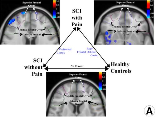
Compared to HCs, SCI + NP demonstrated decreased FC between PAG and right insula, right frontal orbital cortex, right pallidum, dorsal raphe nucleus (DRN), red nuclei (RN), substantia nigra (SN), and ventral posterolateral (VPL) thalamic nuclei. Compared to SCI − NP, SCI + NP demonstrated increased FC between PAG and posterior cingulate cortex (PCC), hippocampus, cerebellar vermis lobules IV and V, and thalamic structures (posterior and lateral pulvinar, the mediodorsal nuclei, and the ventral lateral nuclei). Additionally, decreased FC between the PAG and VPL, geniculate bodies, intralaminar nuclei of thalamus, DRN, RN, SN, and prefrontal cortex was observed in this comparison.

Conclusions

Altered FC between PAG and right anterior insula, VPL, DRN, RN, SN, cerebellar vermis lobules IV and V, frontal cortex, and PCC was associated with NP sequelae of SCI. Additionally, SCI was independently associated with decreased FC between PAG and right posterior insula, cerebellar lobules IV and V, and cerebellar vermis lobules III, IV, and V.
Abstract Image

查看原文
分享 分享
微信好友 朋友圈 QQ好友 复制链接
本刊更多论文
脊髓损伤诱发的神经性疼痛中的uctal灰质周围连通性
背景和目的神经病理性疼痛(NP)是脊髓损伤(SCI)后的一种使人衰弱的症状。脊髓损伤后神经病理性疼痛(Neuropathic pain,NP)是一种使人衰弱的病症。我们的研究采用静息态功能磁共振成像(rsfMRI),旨在证明PAG在SCI后NP中功能连接性(FC)的改变。方法招募了10名SCI患者(SCI + NP,n = 7;SCI - NP,n = 3)和10名健康对照组(HC)。结果与 HCs 相比,SCI + NP 显示 PAG 与右侧岛叶、右侧额眶皮层、右侧苍白球、背侧剑突核(DRN)、红核(RN)、黑质(SN)和丘脑腹后外侧核(VPL)之间的 FC 减少。与 SCI - NP 相比,SCI + NP 显示 PAG 与后扣带回皮层 (PCC)、海马、小脑蚓部第四和第五小叶以及丘脑结构(后部和外侧脉络膜、内侧核和腹侧外侧核)之间的 FC 增加。结论 PAG与右侧前脑岛、VPL、DRN、RN、SN、小脑蚓部小叶IV和V、额叶皮质和PCC之间的FC改变与SCI的NP后遗症有关。此外,SCI还与PAG和右侧后岛叶、小脑第四和第五小叶以及小脑蚓部第三、第四和第五小叶之间的FC下降有关。
本文章由计算机程序翻译,如有差异,请以英文原文为准。
求助全文
约1分钟内获得全文 去求助
来源期刊
Journal of Neuroimaging
Journal of Neuroimaging 医学-核医学
CiteScore
4.70
自引率
0.00%
发文量
117
审稿时长
6-12 weeks
期刊介绍: Start reading the Journal of Neuroimaging to learn the latest neurological imaging techniques. The peer-reviewed research is written in a practical clinical context, giving you the information you need on: MRI CT Carotid Ultrasound and TCD SPECT PET Endovascular Surgical Neuroradiology Functional MRI Xenon CT and other new and upcoming neuroscientific modalities.The Journal of Neuroimaging addresses the full spectrum of human nervous system disease, including stroke, neoplasia, degenerating and demyelinating disease, epilepsy, tumors, lesions, infectious disease, cerebral vascular arterial diseases, toxic-metabolic disease, psychoses, dementias, heredo-familial disease, and trauma.Offering original research, review articles, case reports, neuroimaging CPCs, and evaluations of instruments and technology relevant to the nervous system, the Journal of Neuroimaging focuses on useful clinical developments and applications, tested techniques and interpretations, patient care, diagnostics, and therapeutics. Start reading today!
期刊最新文献
Predicting glioblastoma progression using MR diffusion tensor imaging: A systematic review Nerve cross-sectional area in vincristine-induced polyneuropathy: A nerve ultrasound pilot study Comparison of antithrombogenic coated and uncoated flow diverters in ruptured and unruptured cerebral aneurysms Functional MRI and cognition in multiple sclerosis—Where are we now? Time-dependent MR diffusion analysis of functioning and nonfunctioning pituitary adenomas/pituitary neuroendocrine tumors
×
引用
GB/T 7714-2015
复制
MLA
复制
APA
复制
导出至
BibTeX EndNote RefMan NoteFirst NoteExpress
×
×
提示
您的信息不完整,为了账户安全,请先补充。
现在去补充
×
提示
您因"违规操作"
具体请查看互助需知
我知道了
×
提示
现在去查看 取消
×
提示
确定
0
微信
客服QQ
Book学术公众号 扫码关注我们
反馈
×
意见反馈
请填写您的意见或建议
请填写您的手机或邮箱
已复制链接
已复制链接
快去分享给好友吧!
我知道了
×
扫码分享
扫码分享
Book学术官方微信
Book学术文献互助
Book学术文献互助群
群 号:481959085
Book学术
文献互助 智能选刊 最新文献 互助须知 联系我们:info@booksci.cn
Book学术提供免费学术资源搜索服务,方便国内外学者检索中英文文献。致力于提供最便捷和优质的服务体验。
Copyright © 2023 Book学术 All rights reserved.
ghs 京公网安备 11010802042870号 京ICP备2023020795号-1