Jingyu Zhang, Bin Guo, Yiyi Jiang, Xiaorui Shi, Chong Hu, Zihao Jiao, Fu Wang
{"title":"Bioluminescence and photoacoustic dual-modality imaging of apoptosis using a duramycin-immobilized gold nanorod probe","authors":"Jingyu Zhang, Bin Guo, Yiyi Jiang, Xiaorui Shi, Chong Hu, Zihao Jiao, Fu Wang","doi":"10.1016/j.xcrp.2024.102177","DOIUrl":null,"url":null,"abstract":"<p>Phosphatidylethanolamine (PE) translocation is considered a hallmark event of cellular apoptosis. The development of non-invasive multi-modality probes targeting PE for apoptosis detection holds great promise. Here, we develop a dual-modality imaging probe, duramycin-Fluc-AuNRs (DFA), for detecting apoptosis in tumor cells. DFA is created by linking duramycin peptide and firefly luciferase (Fluc) recombinant protein to gold nanorods (AuNRs). Duramycin exhibits high affinity for PE, while Fluc produces a robust bioluminescence signal, and AuNRs enhance imaging resolution through photoacoustic conversion. The DFA probe demonstrates low toxicity in both cells and mice, showcasing its potential for <em>in vivo</em> applications. In A549 and 4T1 cell lines, the bioluminescence signal of the DFA probe increases with the degree of doxorubicin (Dox)-induced apoptosis. At the mouse level, mice with Dox-triggered apoptosis exhibit higher bioluminescence and photoacoustic imaging signals. Thus, this dual-modality bioluminescence/photoacoustic imaging platform holds significant potential for detecting cellular apoptosis and providing high-performance imaging information.</p>","PeriodicalId":9703,"journal":{"name":"Cell Reports Physical Science","volume":"2 1","pages":""},"PeriodicalIF":7.3000,"publicationDate":"2024-08-29","publicationTypes":"Journal Article","fieldsOfStudy":null,"isOpenAccess":false,"openAccessPdf":"","citationCount":"0","resultStr":null,"platform":"Semanticscholar","paperid":null,"PeriodicalName":"Cell Reports Physical Science","FirstCategoryId":"103","ListUrlMain":"https://doi.org/10.1016/j.xcrp.2024.102177","RegionNum":2,"RegionCategory":"综合性期刊","ArticlePicture":[],"TitleCN":null,"AbstractTextCN":null,"PMCID":null,"EPubDate":"","PubModel":"","JCR":"Q1","JCRName":"CHEMISTRY, MULTIDISCIPLINARY","Score":null,"Total":0}
引用次数: 0
Abstract
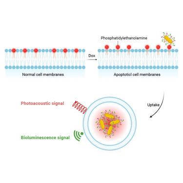
Phosphatidylethanolamine (PE) translocation is considered a hallmark event of cellular apoptosis. The development of non-invasive multi-modality probes targeting PE for apoptosis detection holds great promise. Here, we develop a dual-modality imaging probe, duramycin-Fluc-AuNRs (DFA), for detecting apoptosis in tumor cells. DFA is created by linking duramycin peptide and firefly luciferase (Fluc) recombinant protein to gold nanorods (AuNRs). Duramycin exhibits high affinity for PE, while Fluc produces a robust bioluminescence signal, and AuNRs enhance imaging resolution through photoacoustic conversion. The DFA probe demonstrates low toxicity in both cells and mice, showcasing its potential for in vivo applications. In A549 and 4T1 cell lines, the bioluminescence signal of the DFA probe increases with the degree of doxorubicin (Dox)-induced apoptosis. At the mouse level, mice with Dox-triggered apoptosis exhibit higher bioluminescence and photoacoustic imaging signals. Thus, this dual-modality bioluminescence/photoacoustic imaging platform holds significant potential for detecting cellular apoptosis and providing high-performance imaging information.
期刊介绍:
Cell Reports Physical Science, a premium open-access journal from Cell Press, features high-quality, cutting-edge research spanning the physical sciences. It serves as an open forum fostering collaboration among physical scientists while championing open science principles. Published works must signify significant advancements in fundamental insight or technological applications within fields such as chemistry, physics, materials science, energy science, engineering, and related interdisciplinary studies. In addition to longer articles, the journal considers impactful short-form reports and short reviews covering recent literature in emerging fields. Continually adapting to the evolving open science landscape, the journal reviews its policies to align with community consensus and best practices.


 分享
分享
 求助内容:
求助内容: 应助结果提醒方式:
 扫码关注我们
