Failed endoscopic ultrasound-guided gallbladder drainage across the duodenal covered metallic stent salvaged by using a forward-viewing linear echoendoscope
{"title":"Failed endoscopic ultrasound-guided gallbladder drainage across the duodenal covered metallic stent salvaged by using a forward-viewing linear echoendoscope","authors":"Tesshin Ban, Yoshimasa Kubota, Takashi Joh","doi":"10.1111/den.14931","DOIUrl":null,"url":null,"abstract":"<p>Endoscopic ultrasonography-guided gallbladder drainage (EUS-GBD) has emerged as an alternative to standard percutaneous or transpapillary approaches in fragile patients with acute cholecystitis.<span><sup>1-3</sup></span> Oblique-viewing linear endoscopic ultrasonography (OV-EUS) is used for biliary intervention. However, forward-viewing linear endoscopic ultrasonography (FV-EUS) is applied in certain settings.<span><sup>4, 5</sup></span> Herein, we report salvaged EUS-GBD by using FV-EUS after failure of OV-EUS.</p><p>An 82-year-old man with clinical stage IV pancreatic cancer presented with severe vomiting and initially underwent implantation of a duodenal bulb-covered metallic stent. One week later, this patient underwent endoscopic ultrasonography-guided choledochoduodenostomy due to acute obstructive suppurative cholangitis without intrahepatic biliary dilation (Video S1). One month later, this patient developed antibiotic-refractory acute cholecystitis, which deteriorated into a pericholecystic abscess (Fig. 1). Prioritizing the internal drainage, we attempted EUS-GBD using OV-EUS (EG-580UT; Fujifilm, Tokyo, Japan). The gallbladder was depicted; however, the scope struggled to maneuver in the duodenal metallic stent, and a 19G lancet puncture needle could not advance from the scope channel into the gallbladder (Fig. 2a, Video S1). The following day, we retried EUS-GBD using FV-EUS (TGF-UC260J; Olympus, Tokyo, Japan), which quickly facilitated the gallbladder visualization, needle puncture, 0.025 inch hydrophilic guidewire advancement, electrocautery dilation (Cysto-Gastro-Sets; Endo-flex, Voerde, Germany), and a double-pigtailed plastic stent deployment (Advanix J, 7F, 7 cm; Boston Scientific, Marlborough, MA, USA) (Fig. 2b,c, Video S1). The clinical course was uneventful.</p><p>The maneuverability of the OV-EUS was limited inside the duodenal bulb stent. We needed to down-angle the scope steeply to depict the gallbladder, which obstructed the puncture needle. In this situation, FV-EUS in the long position easily depicted the gallbladder without an angle maneuver. In addition, all the devices showed excellent pushability and trackability, including the puncture needle, dilator, and gallbladder stent, because the target was located vertically in front of the long-positioned FV-EUS.<span><sup>5</sup></span></p><p>Authors declare no conflict of interest for this article.</p>","PeriodicalId":159,"journal":{"name":"Digestive Endoscopy","volume":"36 12","pages":"1389-1390"},"PeriodicalIF":4.7000,"publicationDate":"2024-09-18","publicationTypes":"Journal Article","fieldsOfStudy":null,"isOpenAccess":false,"openAccessPdf":"https://onlinelibrary.wiley.com/doi/epdf/10.1111/den.14931","citationCount":"0","resultStr":null,"platform":"Semanticscholar","paperid":null,"PeriodicalName":"Digestive Endoscopy","FirstCategoryId":"3","ListUrlMain":"https://onlinelibrary.wiley.com/doi/10.1111/den.14931","RegionNum":2,"RegionCategory":"医学","ArticlePicture":[],"TitleCN":null,"AbstractTextCN":null,"PMCID":null,"EPubDate":"","PubModel":"","JCR":"Q1","JCRName":"GASTROENTEROLOGY & HEPATOLOGY","Score":null,"Total":0}
引用次数: 0
Abstract
Endoscopic ultrasonography-guided gallbladder drainage (EUS-GBD) has emerged as an alternative to standard percutaneous or transpapillary approaches in fragile patients with acute cholecystitis.1-3 Oblique-viewing linear endoscopic ultrasonography (OV-EUS) is used for biliary intervention. However, forward-viewing linear endoscopic ultrasonography (FV-EUS) is applied in certain settings.4, 5 Herein, we report salvaged EUS-GBD by using FV-EUS after failure of OV-EUS.
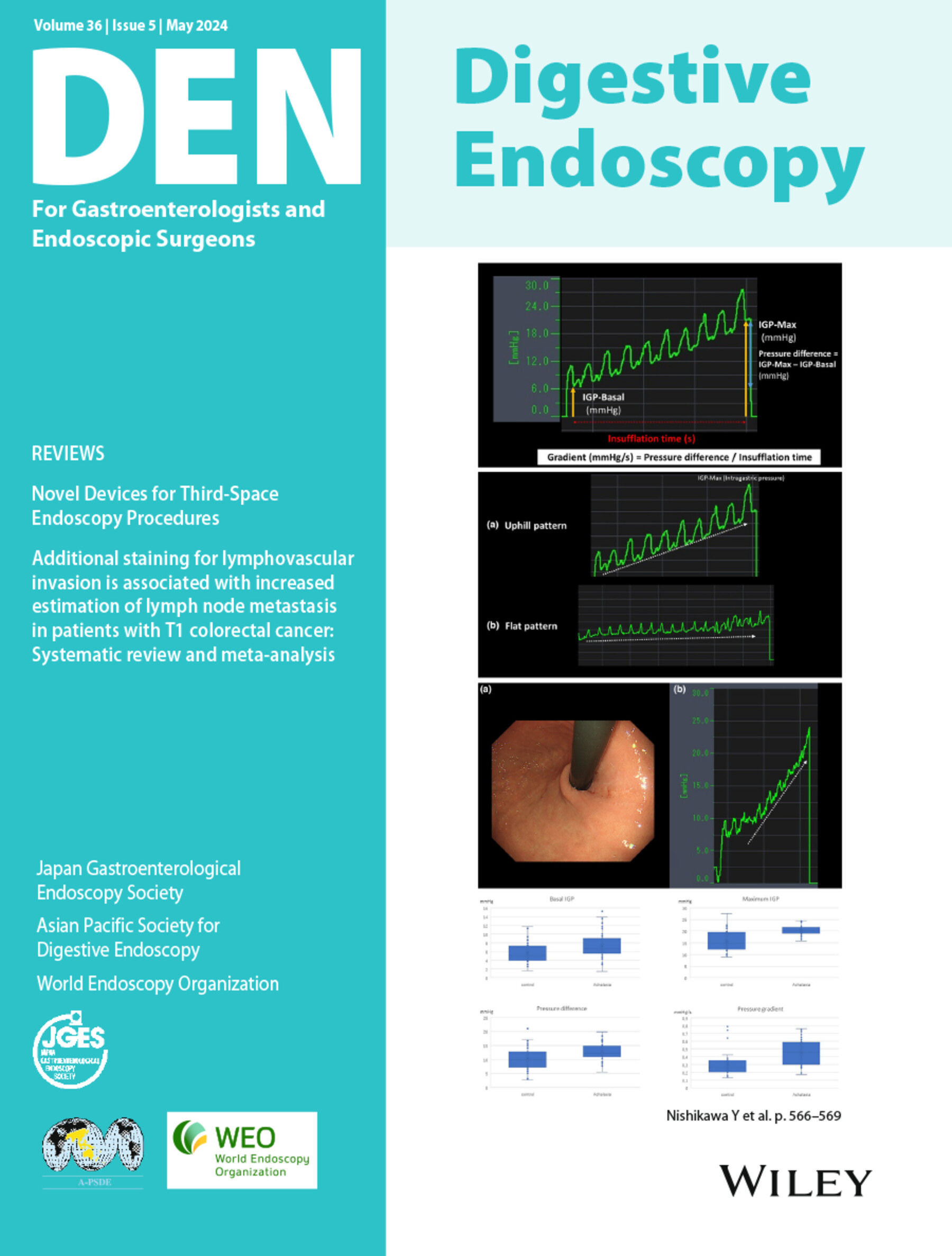
An 82-year-old man with clinical stage IV pancreatic cancer presented with severe vomiting and initially underwent implantation of a duodenal bulb-covered metallic stent. One week later, this patient underwent endoscopic ultrasonography-guided choledochoduodenostomy due to acute obstructive suppurative cholangitis without intrahepatic biliary dilation (Video S1). One month later, this patient developed antibiotic-refractory acute cholecystitis, which deteriorated into a pericholecystic abscess (Fig. 1). Prioritizing the internal drainage, we attempted EUS-GBD using OV-EUS (EG-580UT; Fujifilm, Tokyo, Japan). The gallbladder was depicted; however, the scope struggled to maneuver in the duodenal metallic stent, and a 19G lancet puncture needle could not advance from the scope channel into the gallbladder (Fig. 2a, Video S1). The following day, we retried EUS-GBD using FV-EUS (TGF-UC260J; Olympus, Tokyo, Japan), which quickly facilitated the gallbladder visualization, needle puncture, 0.025 inch hydrophilic guidewire advancement, electrocautery dilation (Cysto-Gastro-Sets; Endo-flex, Voerde, Germany), and a double-pigtailed plastic stent deployment (Advanix J, 7F, 7 cm; Boston Scientific, Marlborough, MA, USA) (Fig. 2b,c, Video S1). The clinical course was uneventful.
The maneuverability of the OV-EUS was limited inside the duodenal bulb stent. We needed to down-angle the scope steeply to depict the gallbladder, which obstructed the puncture needle. In this situation, FV-EUS in the long position easily depicted the gallbladder without an angle maneuver. In addition, all the devices showed excellent pushability and trackability, including the puncture needle, dilator, and gallbladder stent, because the target was located vertically in front of the long-positioned FV-EUS.5
Authors declare no conflict of interest for this article.
期刊介绍:
Digestive Endoscopy (DEN) is the official journal of the Japan Gastroenterological Endoscopy Society, the Asian Pacific Society for Digestive Endoscopy and the World Endoscopy Organization. Digestive Endoscopy serves as a medium for presenting original articles that offer significant contributions to knowledge in the broad field of endoscopy. The Journal also includes Reviews, Original Articles, How I Do It, Case Reports (only of exceptional interest and novelty are accepted), Letters, Techniques and Images, abstracts and news items that may be of interest to endoscopists.


 分享
分享
 求助内容:
求助内容: 应助结果提醒方式:
 扫码关注我们
