Anatomo-functional changes in neural substrates of cognitive memory in developmental amnesia: Insights from automated and manual Magnetic Resonance Imaging examinations
Loïc J. Chareyron, W. K. Kling Chong, Tina Banks, Neil Burgess, Richard C. Saunders, Faraneh Vargha-Khadem
{"title":"Anatomo-functional changes in neural substrates of cognitive memory in developmental amnesia: Insights from automated and manual Magnetic Resonance Imaging examinations","authors":"Loïc J. Chareyron, W. K. Kling Chong, Tina Banks, Neil Burgess, Richard C. Saunders, Faraneh Vargha-Khadem","doi":"10.1002/hipo.23638","DOIUrl":null,"url":null,"abstract":"<p>Despite bilateral hippocampal damage dating to the perinatal or early childhood period and severely impaired episodic memory, patients with developmental amnesia continue to exhibit well-developed semantic memory across the developmental trajectory. Detailed information on the extent and focality of brain damage in these patients is needed to hypothesize about the neural substrate that supports their remarkable capacity for encoding and retrieval of semantic memory. In particular, we need to assess whether the residual hippocampal tissue is involved in this preservation, or whether the surrounding cortical areas reorganize to rescue aspects of these critical cognitive memory processes after early injury. We used voxel-based morphometry (VBM) analysis, automatic (FreeSurfer) and manual segmentation to characterize structural changes in the brain of an exceptionally large cohort of 23 patients with developmental amnesia in comparison with 32 control subjects. Both the VBM and the FreeSurfer analyses revealed severe structural alterations in the hippocampus and thalamus of patients with developmental amnesia. Milder damage was found in the amygdala, caudate, and parahippocampal gyrus. Manual segmentation demonstrated differences in the degree of atrophy of the hippocampal subregions in patients. The level of atrophy in CA-DG subregions and subicular complex was more than 40%, while the atrophy of the uncus was moderate (−24%). Anatomo-functional correlations were observed between the volumes of residual hippocampal subregions in patients and selective aspects of their cognitive performance, viz, intelligence, working memory, and verbal and visuospatial recall. Our findings suggest that in patients with developmental amnesia, cognitive processing is compromised as a function of the extent of atrophy in hippocampal subregions. More severe hippocampal damage may be more likely to promote structural and/or functional reorganization in areas connected to the hippocampus. In this hypothesis, different levels of hippocampal function may be rescued following this variable reorganization. Our findings document not only the extent, but also the limits of circuit reorganization occurring in the young brain after early bilateral hippocampal damage.</p>","PeriodicalId":13171,"journal":{"name":"Hippocampus","volume":"34 11","pages":"645-658"},"PeriodicalIF":2.7000,"publicationDate":"2024-09-13","publicationTypes":"Journal Article","fieldsOfStudy":null,"isOpenAccess":false,"openAccessPdf":"https://onlinelibrary.wiley.com/doi/epdf/10.1002/hipo.23638","citationCount":"0","resultStr":null,"platform":"Semanticscholar","paperid":null,"PeriodicalName":"Hippocampus","FirstCategoryId":"3","ListUrlMain":"https://onlinelibrary.wiley.com/doi/10.1002/hipo.23638","RegionNum":3,"RegionCategory":"医学","ArticlePicture":[],"TitleCN":null,"AbstractTextCN":null,"PMCID":null,"EPubDate":"","PubModel":"","JCR":"Q3","JCRName":"NEUROSCIENCES","Score":null,"Total":0}
引用次数: 0
Abstract
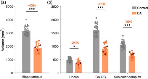
Despite bilateral hippocampal damage dating to the perinatal or early childhood period and severely impaired episodic memory, patients with developmental amnesia continue to exhibit well-developed semantic memory across the developmental trajectory. Detailed information on the extent and focality of brain damage in these patients is needed to hypothesize about the neural substrate that supports their remarkable capacity for encoding and retrieval of semantic memory. In particular, we need to assess whether the residual hippocampal tissue is involved in this preservation, or whether the surrounding cortical areas reorganize to rescue aspects of these critical cognitive memory processes after early injury. We used voxel-based morphometry (VBM) analysis, automatic (FreeSurfer) and manual segmentation to characterize structural changes in the brain of an exceptionally large cohort of 23 patients with developmental amnesia in comparison with 32 control subjects. Both the VBM and the FreeSurfer analyses revealed severe structural alterations in the hippocampus and thalamus of patients with developmental amnesia. Milder damage was found in the amygdala, caudate, and parahippocampal gyrus. Manual segmentation demonstrated differences in the degree of atrophy of the hippocampal subregions in patients. The level of atrophy in CA-DG subregions and subicular complex was more than 40%, while the atrophy of the uncus was moderate (−24%). Anatomo-functional correlations were observed between the volumes of residual hippocampal subregions in patients and selective aspects of their cognitive performance, viz, intelligence, working memory, and verbal and visuospatial recall. Our findings suggest that in patients with developmental amnesia, cognitive processing is compromised as a function of the extent of atrophy in hippocampal subregions. More severe hippocampal damage may be more likely to promote structural and/or functional reorganization in areas connected to the hippocampus. In this hypothesis, different levels of hippocampal function may be rescued following this variable reorganization. Our findings document not only the extent, but also the limits of circuit reorganization occurring in the young brain after early bilateral hippocampal damage.
期刊介绍:
Hippocampus provides a forum for the exchange of current information between investigators interested in the neurobiology of the hippocampal formation and related structures. While the relationships of submitted papers to the hippocampal formation will be evaluated liberally, the substance of appropriate papers should deal with the hippocampal formation per se or with the interaction between the hippocampal formation and other brain regions. The scope of Hippocampus is wide: single and multidisciplinary experimental studies from all fields of basic science, theoretical papers, papers dealing with hippocampal preparations as models for understanding the central nervous system, and clinical studies will be considered for publication. The Editor especially encourages the submission of papers that contribute to a functional understanding of the hippocampal formation.


 分享
分享
 求助内容:
求助内容: 应助结果提醒方式:
 扫码关注我们
