Felix Braczko, Andreas Skyschally, Helmut Lieder, Jakob Nikolas Kather, Petra Kleinbongard, Gerd Heusch
{"title":"Deep learning segmentation model for quantification of infarct size in pigs with myocardial ischemia/reperfusion","authors":"Felix Braczko, Andreas Skyschally, Helmut Lieder, Jakob Nikolas Kather, Petra Kleinbongard, Gerd Heusch","doi":"10.1007/s00395-024-01081-x","DOIUrl":null,"url":null,"abstract":"<p>Infarct size (IS) is the most robust end point for evaluating the success of preclinical studies on cardioprotection. The gold standard for IS quantification in ischemia/reperfusion (I/R) experiments is triphenyl tetrazolium chloride (TTC) staining, typically done manually. This study aimed to determine if automation through deep learning segmentation is a time-saving and valid alternative to standard IS quantification. High-resolution images from TTC-stained, macroscopic heart slices were retrospectively collected from pig experiments (<i>n</i> = 390) with I/R without/with cardioprotection to cover a wide IS range. Existing IS data from pig experiments, quantified using a standard method of manual and subsequent digital labeling of film-scan annotations, were used as reference. To automate the evaluation process with the aim to be more objective and save time, a deep learning pipeline was implemented; the collected images (<i>n</i> = 3869) were pre-processed by cropping and labeled (image annotations). To ensure their usability as training data for a deep learning segmentation model, IS was quantified from image annotations and compared to IS quantified using the existing film-scan annotations. A supervised deep learning segmentation model based on dynamic U-Net architecture was developed and trained. The evaluation of the trained model was performed by fivefold cross-validation (<i>n</i> = 220 experiments) and testing on an independent test set (<i>n</i> = 170 experiments). Performance metrics (Dice similarity coefficient [DSC], pixel accuracy [ACC], average precision [mAP]) were calculated. IS was then quantified from predictions and compared to IS quantified from image annotations (linear regression, Pearson’s <i>r</i>; analysis of covariance; Bland–Altman plots). Performance metrics near 1 indicated a strong model performance on cross-validated data (DSC: 0.90, ACC: 0.98, mAP: 0.90) and on the test set data (DSC: 0.89, ACC: 0.98, mAP: 0.93). IS quantified from predictions correlated well with IS quantified from image annotations in all data sets (cross-validation: <i>r</i> = 0.98; test data set: <i>r</i> = 0.95) and analysis of covariance identified no significant differences. The model reduced the IS quantification time per experiment from approximately 90 min to 20 s. The model was further tested on a preliminary test set from experiments in isolated, saline-perfused rat hearts with regional I/R without/with cardioprotection (<i>n</i> = 27). There was also no significant difference in IS between image annotations and predictions, but the performance on the test set data from rat hearts was lower (DSC: 0.66, ACC: 0.91, mAP: 0.65). IS quantification using a deep learning segmentation model is a valid and time-efficient alternative to manual and subsequent digital labeling.</p>","PeriodicalId":8723,"journal":{"name":"Basic Research in Cardiology","volume":"45 1","pages":""},"PeriodicalIF":8.0000,"publicationDate":"2024-09-30","publicationTypes":"Journal Article","fieldsOfStudy":null,"isOpenAccess":false,"openAccessPdf":"","citationCount":"0","resultStr":null,"platform":"Semanticscholar","paperid":null,"PeriodicalName":"Basic Research in Cardiology","FirstCategoryId":"3","ListUrlMain":"https://doi.org/10.1007/s00395-024-01081-x","RegionNum":1,"RegionCategory":"医学","ArticlePicture":[],"TitleCN":null,"AbstractTextCN":null,"PMCID":null,"EPubDate":"","PubModel":"","JCR":"Q1","JCRName":"CARDIAC & CARDIOVASCULAR SYSTEMS","Score":null,"Total":0}
引用次数: 0
Abstract
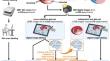
Infarct size (IS) is the most robust end point for evaluating the success of preclinical studies on cardioprotection. The gold standard for IS quantification in ischemia/reperfusion (I/R) experiments is triphenyl tetrazolium chloride (TTC) staining, typically done manually. This study aimed to determine if automation through deep learning segmentation is a time-saving and valid alternative to standard IS quantification. High-resolution images from TTC-stained, macroscopic heart slices were retrospectively collected from pig experiments (n = 390) with I/R without/with cardioprotection to cover a wide IS range. Existing IS data from pig experiments, quantified using a standard method of manual and subsequent digital labeling of film-scan annotations, were used as reference. To automate the evaluation process with the aim to be more objective and save time, a deep learning pipeline was implemented; the collected images (n = 3869) were pre-processed by cropping and labeled (image annotations). To ensure their usability as training data for a deep learning segmentation model, IS was quantified from image annotations and compared to IS quantified using the existing film-scan annotations. A supervised deep learning segmentation model based on dynamic U-Net architecture was developed and trained. The evaluation of the trained model was performed by fivefold cross-validation (n = 220 experiments) and testing on an independent test set (n = 170 experiments). Performance metrics (Dice similarity coefficient [DSC], pixel accuracy [ACC], average precision [mAP]) were calculated. IS was then quantified from predictions and compared to IS quantified from image annotations (linear regression, Pearson’s r; analysis of covariance; Bland–Altman plots). Performance metrics near 1 indicated a strong model performance on cross-validated data (DSC: 0.90, ACC: 0.98, mAP: 0.90) and on the test set data (DSC: 0.89, ACC: 0.98, mAP: 0.93). IS quantified from predictions correlated well with IS quantified from image annotations in all data sets (cross-validation: r = 0.98; test data set: r = 0.95) and analysis of covariance identified no significant differences. The model reduced the IS quantification time per experiment from approximately 90 min to 20 s. The model was further tested on a preliminary test set from experiments in isolated, saline-perfused rat hearts with regional I/R without/with cardioprotection (n = 27). There was also no significant difference in IS between image annotations and predictions, but the performance on the test set data from rat hearts was lower (DSC: 0.66, ACC: 0.91, mAP: 0.65). IS quantification using a deep learning segmentation model is a valid and time-efficient alternative to manual and subsequent digital labeling.
期刊介绍:
Basic Research in Cardiology is an international journal for cardiovascular research. It provides a forum for original and review articles related to experimental cardiology that meet its stringent scientific standards.
Basic Research in Cardiology regularly receives articles from the fields of
- Molecular and Cellular Biology
- Biochemistry
- Biophysics
- Pharmacology
- Physiology and Pathology
- Clinical Cardiology


 分享
分享
 求助内容:
求助内容: 应助结果提醒方式:
 扫码关注我们
