Viola Katharina Vetter, Martina Haberecker, Florian Alexander Huber, Chantal Pauli
{"title":"Aberrant positivity for BCOR immunohistochemistry in merkel cell carcinoma - a potential diagnostic pitfall.","authors":"Viola Katharina Vetter, Martina Haberecker, Florian Alexander Huber, Chantal Pauli","doi":"10.1186/s13000-024-01552-8","DOIUrl":null,"url":null,"abstract":"<p><strong>Backrgound: </strong>Merkel cell carcinoma (MCC) is a rare, aggressive primary cutaneous neuroendocrine carcinoma, frequently associated with clonal Merkel cell polyomavirus integration. MCC can pose significant diagnostic challenges due to its diverse clinical presentation and its broad histological differential diagnosis. Histologically, MCC presents as a small-round-blue cell neoplasm, where the differential diagnosis includes basal cell carcinoma, melanoma, hematologic malignancies, round cell sarcoma and metastatic small cell carcinoma of any site. We here report strong aberrant immunoreactivity for BCOR in MCC, a marker commonly used to identify round cell sarcomas and other neoplasms with BCOR alterations.</p><p><strong>Methods: </strong>Based on strong BCOR expression in three index cases of MCC, clinically mistaken as sarcoma, a retrospective analysis of three patient cohorts, comprising 31 MCC, 19 small cell lung carcinoma (SCLC) and 5 cases of neoplasms with molecularly confirmed BCOR alteration was conducted. Immunohistochemical staining intensity and localization for BCOR was semi-quantitatively analyzed.</p><p><strong>Results: </strong>Three cases, clinically and radiologically mimicking a sarcoma were analyzed in our soft tissue and bone pathology service. Histologically, the cases showed sheets of a small round blue cell neoplasm. A broad panel of immunohistochemistry was used for lineage classification. Positivity for synaptophysin, CK20 and Merkel cell polyoma virus large T-antigen lead to the diagnosis of a MCC. Interestingly, all cases showed strong positive nuclear staining for BCOR, which was included for the initial work-up with the clinical differential of a round cell sarcoma. We analyzed a larger retrospective MCC cohort and found aberrant weak to strong BCOR positivity (nuclear and/or cytoplasmic) in up to 90% of the cases. As a positive control, we compared the expression to a small group of BCOR-altered neoplasms. Furthermore, we investigated a cohort of SCLC as another neuroendocrine neoplasm and found in all cases a diffuse moderate to strong BCOR positivity.</p><p><strong>Conclusions: </strong>This study demonstrates that neuroendocrine neoplasms, such as MCC and SCLC can express strong aberrant BCOR. This might represent a diagnostic challenge or pitfall, in particular when MCC is clinically mistaken as a soft tissue or a bone sarcoma.</p>","PeriodicalId":11237,"journal":{"name":"Diagnostic Pathology","volume":"19 1","pages":"130"},"PeriodicalIF":2.3000,"publicationDate":"2024-09-27","publicationTypes":"Journal Article","fieldsOfStudy":null,"isOpenAccess":false,"openAccessPdf":"https://www.ncbi.nlm.nih.gov/pmc/articles/PMC11437883/pdf/","citationCount":"0","resultStr":null,"platform":"Semanticscholar","paperid":null,"PeriodicalName":"Diagnostic Pathology","FirstCategoryId":"3","ListUrlMain":"https://doi.org/10.1186/s13000-024-01552-8","RegionNum":3,"RegionCategory":"医学","ArticlePicture":[],"TitleCN":null,"AbstractTextCN":null,"PMCID":null,"EPubDate":"","PubModel":"","JCR":"Q2","JCRName":"PATHOLOGY","Score":null,"Total":0}
引用次数: 0
Abstract
Backrgound: Merkel cell carcinoma (MCC) is a rare, aggressive primary cutaneous neuroendocrine carcinoma, frequently associated with clonal Merkel cell polyomavirus integration. MCC can pose significant diagnostic challenges due to its diverse clinical presentation and its broad histological differential diagnosis. Histologically, MCC presents as a small-round-blue cell neoplasm, where the differential diagnosis includes basal cell carcinoma, melanoma, hematologic malignancies, round cell sarcoma and metastatic small cell carcinoma of any site. We here report strong aberrant immunoreactivity for BCOR in MCC, a marker commonly used to identify round cell sarcomas and other neoplasms with BCOR alterations.
Methods: Based on strong BCOR expression in three index cases of MCC, clinically mistaken as sarcoma, a retrospective analysis of three patient cohorts, comprising 31 MCC, 19 small cell lung carcinoma (SCLC) and 5 cases of neoplasms with molecularly confirmed BCOR alteration was conducted. Immunohistochemical staining intensity and localization for BCOR was semi-quantitatively analyzed.
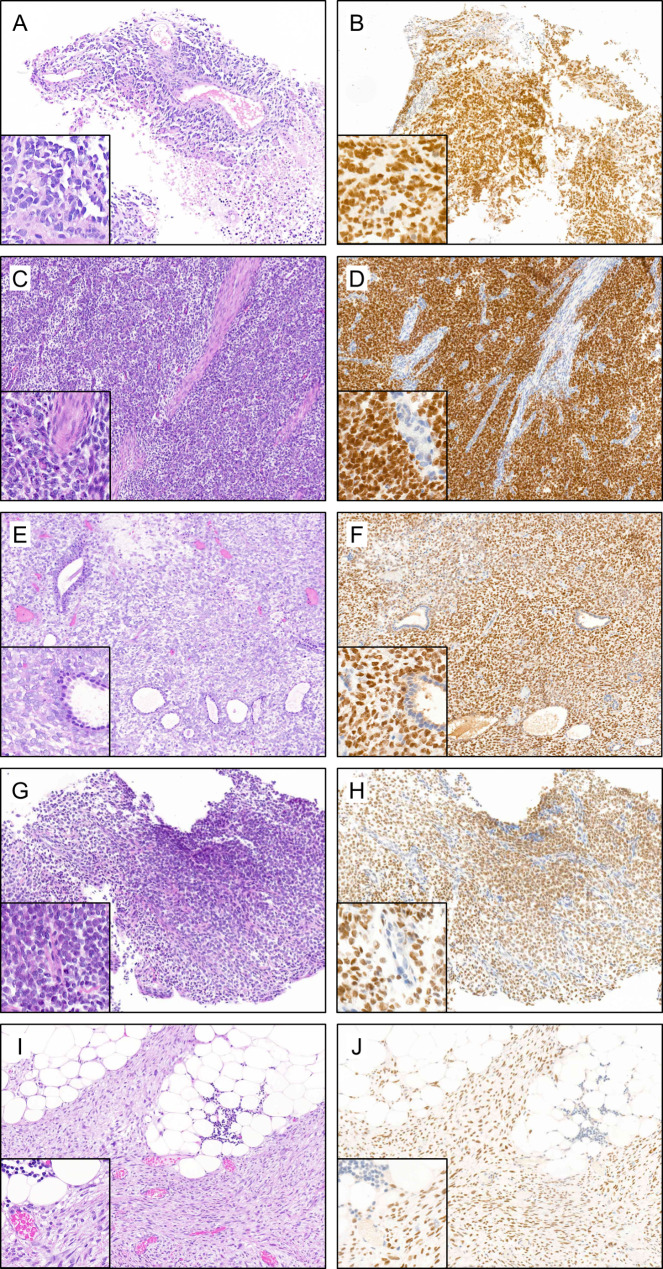
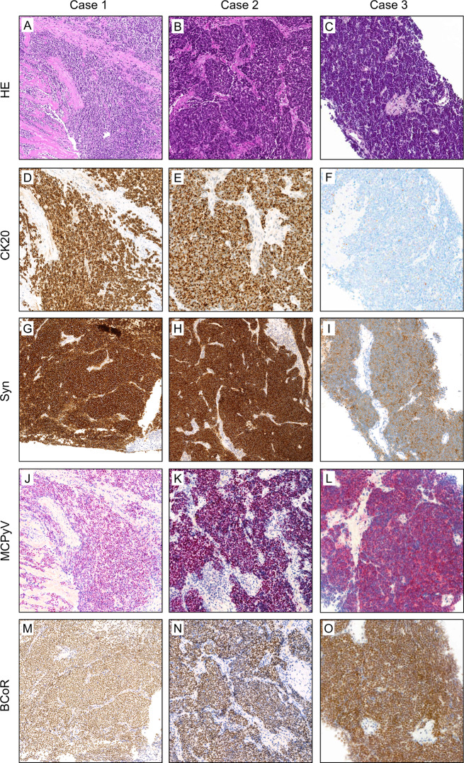
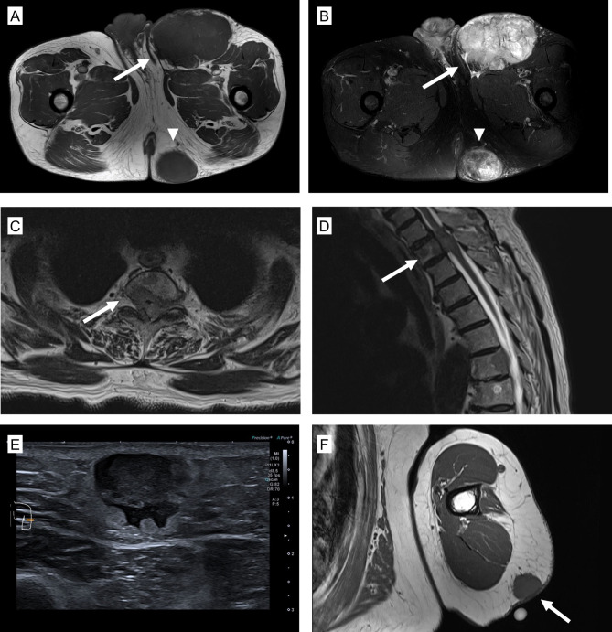
Results: Three cases, clinically and radiologically mimicking a sarcoma were analyzed in our soft tissue and bone pathology service. Histologically, the cases showed sheets of a small round blue cell neoplasm. A broad panel of immunohistochemistry was used for lineage classification. Positivity for synaptophysin, CK20 and Merkel cell polyoma virus large T-antigen lead to the diagnosis of a MCC. Interestingly, all cases showed strong positive nuclear staining for BCOR, which was included for the initial work-up with the clinical differential of a round cell sarcoma. We analyzed a larger retrospective MCC cohort and found aberrant weak to strong BCOR positivity (nuclear and/or cytoplasmic) in up to 90% of the cases. As a positive control, we compared the expression to a small group of BCOR-altered neoplasms. Furthermore, we investigated a cohort of SCLC as another neuroendocrine neoplasm and found in all cases a diffuse moderate to strong BCOR positivity.
Conclusions: This study demonstrates that neuroendocrine neoplasms, such as MCC and SCLC can express strong aberrant BCOR. This might represent a diagnostic challenge or pitfall, in particular when MCC is clinically mistaken as a soft tissue or a bone sarcoma.
期刊介绍:
Diagnostic Pathology is an open access, peer-reviewed, online journal that considers research in surgical and clinical pathology, immunology, and biology, with a special focus on cutting-edge approaches in diagnostic pathology and tissue-based therapy. The journal covers all aspects of surgical pathology, including classic diagnostic pathology, prognosis-related diagnosis (tumor stages, prognosis markers, such as MIB-percentage, hormone receptors, etc.), and therapy-related findings. The journal also focuses on the technological aspects of pathology, including molecular biology techniques, morphometry aspects (stereology, DNA analysis, syntactic structure analysis), communication aspects (telecommunication, virtual microscopy, virtual pathology institutions, etc.), and electronic education and quality assurance (for example interactive publication, on-line references with automated updating, etc.).


 分享
分享
 求助内容:
求助内容: 应助结果提醒方式:
 扫码关注我们
